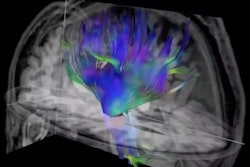
Anesthesia can have negative effects on cognitive function in patients who receive it. In a new study, researchers used diffusion-tensor (DTI)-MRI to shed some light on why, according to a study published March 26 in Plos One.
The results highlight the need for more research on the connection between anesthesia and changes in the brain, wrote a team led by Cheuk Tang, PhD, of Icahn School of Medicine at Mount Sinai, New York City.
"[Further] investigations into whether these changes in white matter microstructure are related to loss of consciousness are warranted," the group wrote.
In older patients, cognitive dysfunction after surgery with general anesthesia is a well-known problem, but the effects of anesthesia on brain structure are unclear, the authors noted.
"Cognitive dysfunction in elderly patients after they undergo surgery under general anesthesia is a well-known phenomenon, but it is unclear whether this is caused by the surgical procedures or the anesthesia," they wrote. "Little is known about how general anesthesia causes a reduction in nerve transmission and subsequent loss of consciousness and the effects of general anesthesia on brain structure has not been studied before."
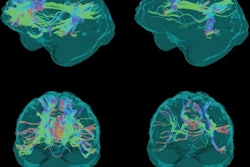
Under the auspices of a larger study called Trajectory of Recovery in the Elderly (TORIE), which explored the effects of anesthesia on cognitive function in 72 elderly patients in the absence of surgery, Tang and colleagues performed an analysis of the effects of general anesthesia on brain white matter using DTI-MRI and calculating various metrics such as fractional anisotropy, radial diffusivity, axial diffusivity, and mean diffusivity maps.
Study participants underwent five MRI scans: one before receiving the anesthesia sevoflurane; two during a two-hour period of anesthesia (one at 40 minutes, another at 100 minutes), and two more one day after anesthesia and seven days after. The patients underwent these exams in an imaging suite that had an MRI-compatible anesthesia setup and set of vital sign monitors.
The researchers noted temporary changes in the DTI metrics when they compared pre- and postanesthesia MRIs: Fractional anisotropy decreased during the time patients were anesthetized, while mean diffusivity, radial diffusivity, and axial diffusivity all increased.
The authors offered the following hypotheses for causes of these changes:
- Glial cell shrinkage due to reduced activity
- The glymphatic system
- The microtubule system
- Intracranial pressure
Even though the effect of anesthesia on the brain's white matter appears to be temporary, more research should be conducted, according to the investigators.
"[It] is important to consider these results in research studies that necessitate the use of anesthesia, given that general anesthesia can produce significant changes in diffusion measures of white matter integrity as well as in volume of specific brain regions," they concluded.