Artificial intelligence (AI)-based image reconstruction enables significantly faster brain MRI scan times while maintaining quantification accuracy and enhancing image quality, according to research from imaging services provider RadNet.
Researchers led by neuroradiologist Dr. Suzie Bash performed a prospective, multicenter study to assess the benefits of utilizing AI-based reconstruction of 60% accelerated MRI with quantitative volumetric MR image analysis. They found that the combination offered a significant improvement over traditional MRI acquisition methods.
"Shorter scan times may boost utilization of volumetric quantitative MRI in routine clinical settings," said Bash, who presented the findings at ECR 2021.
Deep learning can offer a number of benefits in image reconstruction, including superior image quality, higher perceived signal-to-noise ratio, and higher perceived imaging contrast, according to Bash. It can also reduce artifacts and yield higher perceived spatial resolution, she said.
"Our question was whether it's possible to effectively apply more than one AI solution to obtain the benefits of both tools," Bash said. "Specifically, our goal was to apply an image reconstruction deep-learning tool to obtain faster MRIs, while still maintaining quantitative accuracy of the datasets."
The researchers prospectively recruited 40 subjects who underwent clinical brain MRI exams on six scanners from five different institutions. The participants included cognitively normal subjects as well as patients with mild cognitive impairment (MCI) and Alzheimer's disease.
Two T1-weighted volumetric scans were acquired: one using the routine clinical protocol -- considered to be the standard of care -- and the other at 60% faster acquisition speed. After the accelerated exams were enhanced by image reconstruction software (SubtleMR, Subtle Medical), automatic brain lesion quantification software was applied (NeuroQuant, CorTechs Lab) to both the enhanced accelerated images and the standard-of-care images.
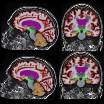
![Representative 3D T1-weighted multiplanar images with volumetric segmentation on a 3-tesla scanner. [Left to right]: Sagittal, coronal, and axial T1-weighted images with standard protocols (scan time, five minutes and 44 seconds) on the top row and deep learning-enhanced accelerated imaging (scan time, two minutes and 18 seconds) on bottom row. Images courtesy of Dr. Suzie Bash.](https://img.auntminnie.com/files/base/smg/all/image/2021/03/am.2021_03_15_17_22_4193_2021_03_17_AV_Insider_redo.png?auto=format%2Ccompress&dpr=2&fit=max&q=70&w=700) Representative 3D T1-weighted multiplanar images with volumetric segmentation on a 3-tesla scanner. [Left to right]: Sagittal, coronal, and axial T1-weighted images with standard protocols (scan time, five minutes and 44 seconds) on the top row and deep learning-enhanced accelerated imaging (scan time, two minutes and 18 seconds) on bottom row. Images courtesy of Dr. Suzie Bash.
Representative 3D T1-weighted multiplanar images with volumetric segmentation on a 3-tesla scanner. [Left to right]: Sagittal, coronal, and axial T1-weighted images with standard protocols (scan time, five minutes and 44 seconds) on the top row and deep learning-enhanced accelerated imaging (scan time, two minutes and 18 seconds) on bottom row. Images courtesy of Dr. Suzie Bash.Next, a neuroradiologist classified the cases into clinical disease categories (normal/MCI or Alzheimer's disease), Bash said. The concordance of image biomarkers -- hippocampal occupancy score, hippocampi volume, superior lateral ventricles volume, and inferior lateral ventricles volume -- was also assessed for both sets of images.
To evaluate image quality, the researchers also presented blinded, randomized, side-by-side multiplanar datasets to two neuroradiologists. These readers rated 360 image series on a 5-point Likert scale for six features: apparent signal-to-noise ratio, image sharpness, artifacts, lesion conspicuity, image contrast, and gray-white differentiation.
Despite a 60% shorter scan time, the deep learning-enhanced images were statistically superior (p < 0.05) for perceived quality across all imaging features, according to Bash. Furthermore, linear regression analysis showed that the methods had very high correlation for quantitative volumetric biomarkers:
- Hippocampal occupancy score: r² = 0.9953
- Hippocampi volume: r² = 0.9732
- Superior lateral ventricles volume: r² = 0.9977
- Inferior lateral ventricles volume: r² = 0.992
Furthermore, the researchers found no difference in clinical classification between the standard-of-care MR images and the deep learning-enhanced accelerated images.
"This trial supports the reliability, efficiency, and clinical utility of deep learning-based enhancement for quantitative imaging," Bash said.