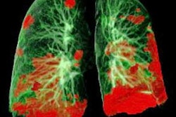
A machine-learning algorithm based on radiomics features of pure ground-glass nodules on CT can yield a promising level of accuracy for differentiating noninvasive from invasive pulmonary adenocarcinoma prior to surgery, according to research published online November 10 in Clinical Radiology.
A team of researchers led by J. Cai of Dalian Medical University in Dalian, China, trained a support vector machine (SVM) machine-learning model to distinguish pulmonary adenocarcinoma in situ or minimally invasive adenocarcinoma from invasive adenocarcinoma using six CT radiomics features. In a retrospective study, the group found that the algorithm achieved an accuracy of 90.2% on a test set, outperforming two radiologists.
"This model could assist radiologists in predicting invasiveness and guide clinicians when choosing the optimal therapeutic strategy," the authors wrote.
As the different subtypes of lung adenocarcinoma have different prognostic and treatment outcomes, it would be helpful for thoracic surgeons in surgical decision making if radiologists could accurately and before surgery distinguish between noninvasive and invasive pulmonary adenocarcinoma. However, visual radiographic features on CT lack the specificity needed for radiologists to perform that task in clinical practice, according to the researchers.
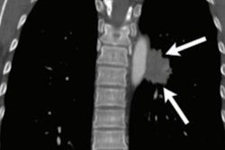
As a result, the team sought to utilize radiomics and machine learning to develop a noninvasive preoperative tool for assessing the invasiveness of pure ground-glass nodules. The group included 136 lung adenocarcinoma patients from December 2016 to September 2019 in their study, including 37 males and 99 females with an average age of 52.9 ± 10.9 years. Of these patients, 39 were histopathologically diagnosed with invasive adenocarcinoma and 97 had noninvasive lesions, including 15 with adenocarcinoma in situ and 82 with minimally invasive adenocarcinoma.
The researchers then trained an SVM machine-learning model using 95 pure ground-glass nodules, including 27 cases of invasive adenocarcinoma and 68 cases of adenocarcinoma in situ or minimally invasive adenocarcinoma. After radiomics features were extracted from manual 3D segmentation of unenhanced CT images from the patient's last preoperative CT exam, Cai and colleagues found that six features yielded the best performance for the machine-learning model.
Next, they evaluated the final model on a separate test set of 41 nodules, consisting of 12 cases of invasive adenocarcinoma and 29 cases of adenocarcinoma in situ or minimally invasive adenocarcinoma. They also compared the performance of the algorithm on the test set with that of two radiologists, one with 10 years of experience and the other with 20 years of experience.
| AI performance for distinguishing between adenocarcinoma in situ/minimally invasive carcinoma from invasive adenocarcinoma appearing as pure ground-glass nodules | |||
| Radiologist 1 (10 years of experience) | Radiologist 2 (20 years of experience) | Radiomics-based machine-learning model | |
| Sensitivity | 50% | 75% | 91.7% |
| Specificity | 86.2% | 82.8% | 89.7% |
| Accuracy | 75.6% | 80.5% | 90.2% |
"Although the false-positive rates of the two radiologists were close to the model, the sensitivity and accuracy were noticeably lower than those in the current study," the authors wrote.
In future work, the researchers noted that other machine-learning classifiers should be compared with their SVM model in larger datasets. Also, the model should be utilized with data from other institutions in order to evaluate its generalizability, according to the authors.