Researchers have found that nonchest CT exams taken in the emergency room for other indications may also capture pulmonary findings suspicious for COVID-19, according to a study published May 11 in Radiology.
The study results suggest that radiologists must keep in mind that patients with COVID-19 can initially present with symptoms other than respiratory ones but still have the disease, wrote a team led by Dr. Rydhwana Hossain of the University of Maryland School of Medicine in Baltimore.
"In addition to pulmonary manifestations, patients with COVID-19 may present with ... neurological and gastrointestinal symptoms," the team wrote. "Patients who present solely with gastrointestinal symptoms are more likely to be undiagnosed [for COVID-19] and remain in their community posing a continued infectious risk."
Patients who come to the emergency room because of gastrointestinal or neurological symptoms but who are not known to be positive for COVID-19 may undergo CT imaging that includes portions of the lung, the team noted. Hossain and colleagues investigated cases from 119 patients who had nonrespiratory CT for gastrointestinal or neurological symptoms and were noted to have findings suspicious for COVID-19.
The team included data collected between March 10 and April 6 from three institutions, two of which were in New York City, at the time considered a "hotspot" for COVID-19. Sixty-two of the 119 patients (group 1) had a reverse transcriptase polymerase chain reaction (RT-PCR) assay before the CT scan because they presented in the emergency room with cough and fever and COVID-19 was suspected, while 57 patients (group 2) had RT-PCR after the CT exam due to incidental abnormal pulmonary findings.
Of the 119 patients, 101 had abdomen/pelvis CT and 18 had cervical spine/neck CT. The researchers found the following:
- In 52% of group 1 patients, nonchest CT findings showed evidence of COVID-19 pneumonia.
- In 77% of group 2 patients, the findings showed COVID-19 evidence.
- The most common CT lung findings were ground-glass opacity (96%) and consolidation (40%).
- Of the total patient cohort, 24% required vasopressor medication or intubation, and 23% died.
- Those who had cervical spine/neck CT had worse outcomes than those who underwent abdominal/pelvic CT (p=0.01).
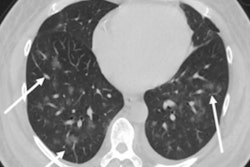
 A 33-year-old man presenting with right lower quadrant abdominal pain, found to have acute appendicitis on abdominal/pelvic CT. Axial (left) and coronal (right) views on lung windows demonstrate focal peripheral ground-glass opacity in the right lung base. Images courtesy of the RSNA.
The study findings confirm that it's important to keep COVID-19 disease in mind as a diagnostic possibility, even if patients are presenting without typical symptoms, the authors concluded.
"Radiologists should maintain a high index of suspicion with respect to the lungs in patients with primary extrapulmonary clinical symptoms who undergo nonchest CT studies to facilitate earlier diagnosis of COVID-19 related pneumonia," the team wrote.