An advanced imaging technique called black-blood CT can leverage cinematic rendering technology to provide a detailed view inside the lumen of vasculature and potentially bolster cardiac evaluation, according to an article published online October 1 in the Journal of Cardiovascular Computed Tomography.
Researchers from Johns Hopkins University developed the black-blood CT method in an effort to enhance the visualization of cardiac anatomy and pathology using cinematic rendering software readily available at their institution. Early clinical applications of the technique using contrast-enhanced chest CT scans have proved promising for various scenarios, especially in demonstrating findings within the lumen of cardiac vasculature.
"The black-blood [CT] method appears to provide high levels of intraluminal anatomic detail for cardiac CT imaging," Dr. Steven Rowe, PhD; Dr. Elliot Fishman; and colleagues wrote. "This may facilitate the detection of important pathologic entities such as intramural thrombus, and may also allow improved evaluation of cardiac devices."
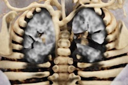
 Black-blood CT showing a calcified mitral annulus. The image was generated using cinematic rendering technology. All images courtesy of Dr. Elliot Fishman.
The color of blood
Advances in CT technology have paved the way for the clinical use of cinematic rendering over the past few years, and a growing number of institutions are beginning to explore its potential advantages in diagnostic imaging. Various groups have confirmed the technique's clinical utility in a wide range of settings, from musculoskeletal trauma imaging to diagnosing colitis.
Cinematic rendering employs a global lighting model to add depth and shadowing effects to volumetric CT datasets, substantially increasing their photorealism beyond the capabilities of traditional volume rendering.
With cinematic rendering software (syngo.via VB30, Siemens Healthineers) already integrated into the CT workflow at their institution, Rowe, Fishman, and colleagues have been creating cinematic rendering images on a daily basis for a variety of clinical scenarios.
They recently developed a preset to visualize intraluminal structures in the heart that were initially acquired on CT scans. This preset serves as the starting point from which radiologists are able to make subtle adjustments to window width and level settings before generating the final image. The total amount of time required to create the cinematic rendering images is typically five minutes, with less than a minute of that time spent on manual adjustments.
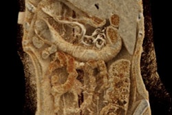
 Black-blood CT displaying a calcified aortic valve.
The group applied the technique to several contrast-enhanced chest CT datasets and found it produced cinematic rendering images that displayed blood in black, a visual effect resembling black-blood MRI.
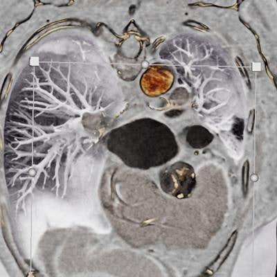
Intraluminal view
In the current study, the researchers investigated the possible advantages of black-blood CT over conventional CT and volume rendering for several cases, including the following:
- A 71-year-old woman with an ascending aortic aneurysm: The blood pool in the patient's 2D CT scans appeared overexposed and obscured the surface detail of the underlying cardiac chamber. In contrast, by making the blood appear black, black-blood CT allowed for better visualization of the interior heart wall. This, in turn, revealed myocardial trabeculation (i.e., thinning) in the left ventricle and anterolateral papillary muscle.
- A 51-year-old woman with embolic renal infarcts: The patient presented with abdominal pain, and conventional CT showed she had embolic renal infarcts. The embolic renal infarcts were clearly visible to radiologists using any of the techniques, but black-blood CT greatly improved visualization of the pulmonary vasculature, which could benefit cases involving complications in that area.
- An 80-year-old man with prior transcatheter arterial valve replacement: Both volume rendering and black-blood CT showed myocardial trabeculation in the left ventricle of the patient that was difficult to appreciate on conventional CT. However, black-blood CT offered a much more detailed view of the metallic stent inside the patient's aorta, compared with the volume-rendered scans.
Overall, cinematic rendering demonstrated utility in multiple facets of cardiac CT, the authors noted. The technique was particularly helpful in identifying changes in myocardial trabeculation, depicting prosthetic valves and other devices, diagnosing vegetations, and providing intraluminal views of aneurysms. Yet black-blood CT also faces the possibility of obscuring some pathology due to shadowing effects, potentially limiting its application in certain cases.
"There appears to be significant promise in utilizing black-blood [CT] for cardiac intraluminal visualization. ... However, important aspects of this methodology remain to be studied," they wrote.