In life, timing is everything. With sodium fluoride (NaF) PET, interpreting results three hours after tracer injection instead of the customary one-hour delay improved image quality and standardized uptake values (SUVs), leading to significantly better coronary plaque detection, according to a September 13 article in the Journal of Nuclear Medicine.
On average, the extra two hours of wait time increased corrected maximum SUV (SUVmax) results by 92% (range: 33%-225%) and target-to-background ratios by 80% (range: 20%-177%). With these changes, seven more lesions were identified at the three-hour mark than one hour after NaF injection.
"Our findings have important clinical implications. ... By decreasing the background (blood pool) signal, not only do the target-to-background values improve, but also lesions which were difficult to detect one hour postinjection can be easily identified," wrote the researchers led by Dr. Jacek Kwiecinski from Cedars-Sinai Medical Center.
Clinicians have become accustomed to analyzing increased NaF uptake with PET and coronary CT angiography (CCTA) scans one hour after radiopharmaceutical injection to detect plaque rupture in patients with acute myocardial infarction, as well as in people with stable coronary artery disease but who have high-risk features on intravascular ultrasound.
While semiquantitative analysis is "feasible with one-hour images, it can be difficult to discriminate plaques with NaF uptake from noise," the researchers wrote. Recent studies have also suggested that the "optimal time for atherosclerotic plaque imaging with sodium fluoride might differ from the one-hour postinjection time point used for bone imaging."
To investigate this question further, Kwiecinski and colleagues analyzed 20 patients (mean age, 67 ± 7 years) who had stable coronary artery disease. All subjects underwent CCTA and NaF-PET/CT (Discovery 710, GE Healthcare) with a dose of 250 MBq of NaF.
One hour after PET imaging, contrast-enhanced CCTA was performed with prospective electrocardiogram (ECG) triggering if the heart rate was less than 60 beats per minute and regular or ECG-gated helical acquisition if the heart rate was more than 60 beats per minute or irregular.
A second low-dose CT scan was performed for attenuation correction, with NaF-PET performed three hours after tracer injection using the same protocol as the one-hour study. After one hour, 26 CCTA segments were deemed positive for NaF uptake, 11 (41%) of which were in the left anterior descending artery.
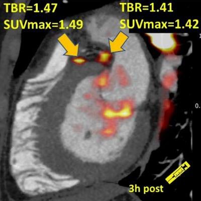
 Images show significant NaF uptake in proximal left anterior descending, proximal circumflex (A), and distal right coronary artery (B) plaques, which had a target-to-background ratio (TBR) of less than 1.0 on one-hour PET and showed uptake exceeding the 1.25 TBR threshold at three hours. Image courtesy of JNM.
At three hours, NaF activity was "markedly reduced, and coronary lesions were more conspicuous," the authors wrote. All previously identified 26 segments were seen, as well as seven additional CTA segments that had been negative for NaF uptake after one hour.
Despite the small cohort of only 20 patients, they noted that their "findings are supported by statistically significant data."
"A greater than a one-hour delay may improve the detection of NaF uptake in coronary artery plaques," the group concluded.