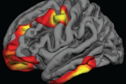
Fetal MRI brain scans can advance the detection of abnormalities due to severe fetal anemia and correlate subsequent deficiencies with poor neurological development, according to a French study published online June 6 in the European Journal of Obstetrics and Gynecology and Reproductive Biology.
Among some two dozen pregnant mothers, MRI also detected brain lesions that were missed on initial ultrasound scans, which are part of the standard follow-up after intrauterine transfusion (IUT) to bring red blood cell counts back to normal.
"Our results indicate that fetal brain MRI makes it possible to better detect brain lesions in the management of fetal anemia requiring IUT and to confirm the severity of certain lesions seen on ultrasound," wrote lead author Dr. Louise Ghesquière and colleagues from Jeanne de Flandre Hospital in Lille. "Fetal brain MRI appears to be particularly important in the follow-up of fetal anemia of acute mechanism."
Standard of care
Intrauterine transfusion is a procedure in which red blood cells are injected into a pregnant woman who has severe acute fetal anemia. The procedure is intended to maintain the mother-to-be's immune system until the baby's birth. Left untreated, severe anemia can cause a variety of health issues, including fetal-maternal hemorrhage and/or a sudden decrease in blood flow.
It's been estimated that IUT can increase survival rates by as much as 90%, except when complicated by severe swelling of the fetus, also known as hydrops fetalis. However, even when newborns survive fetal anemia, the condition often causes brain lesions and related cerebral damage that, in turn, can stunt a baby's development.
Ultrasound has been used to detect signs of recurrent anemia, as well as ischemic or hemorrhagic brain damage, but the modality cannot reliably determine a connection between the abnormalities and a child's neurological development. A 2017 study by Griffiths et al gave credence to in utero MRI, which showed improved diagnostic accuracy in visualizing fetal brain abnormalities. The researchers also recommended MRI as an adjunct to ultrasound to better evaluate and manage the health of the fetus and mother.
With those considerations in mind, Ghesquière and colleagues sought to "investigate the contribution of fetal brain MRI to the diagnosis of cerebral abnormalities in cases of fetal anemia requiring IUT," they wrote.
Assessing anemia
The retrospective, single-center study included 89 expectant mothers seen between 2005 and 2016 who had a variety of fetal anemic conditions that required IUT. The indications for follow-up MRI included hydrops fetalis, more than three IUTs, severe acute anemia, and/or an abnormality seen on ultrasound (Eur J Obstet Gynecol Reprod Biol, June 6, 2018).
Clinicians performed ultrasound exams (Voluson E8 or E10, GE Healthcare) on all 89 patients. Of those women, 28 (32%) underwent a 1.5- or 3-tesla fetal MRI brain scan (Achieva, Philips Healthcare) the following week. The researchers also noted subjects' obstetric histories, the etiology and severity of the fetal anemia, any complications, and related IUT information.
The mean gestational age at the time of the first fetal brain MRI scan was 31.2 weeks (± 1.8 weeks). Four patients (14%) underwent two fetal MRI brain scans after clinicians detected abnormalities on the first scan.
Of the 28 patients who had an MRI, 12 (43%) presented with abnormalities as a result of the fetal anemia. The anomalies were visible only on MRI in 10 cases (36%) and on both MRI and ultrasound in two patients (7%). In addition, an abnormal MRI result contradicted normal ultrasound findings in five cases (18%).
MRI's efficacy
Seven cases (58%) among the 12 abnormal fetal MRI results had poor neurological prognoses: Two women chose to medically terminate their pregnancies, two children had severe developmental delays, and three children eventually had difficulty in school. The other five children (42%) received favorable neurological prognoses.
"In our study ... we found fetal brain MRI to be a useful additional tool in the prenatal detection of brain lesions in the follow-up of severe anemia," the authors concluded. "MRI revealed some brain lesions that were not visible on ultrasound."
MR imaging may be particularly beneficial because intrauterine transfusions have improved the prognosis for patients with fetal anemia, Ghesquière and colleagues added.
"The main concern in the management of this condition has become child neurological development," they wrote. "Since prenatal counseling is based on the interpretation of imaging, there is a place for fetal brain MRI just as it is used in other pathologies."