The ability to detect cancerous gastrointestinal and pancreatic neuroendocrine tumors (NETs) soon might get a boost from a radiolabeled PET tracer that is better able to bind to tumors, according to a study published in the June issue of the Journal of Nuclear Medicine.
Swiss and German researchers are developing gallium-68 (Ga-68) OPS202, a somatostatin receptor subtype 2 (sst2) antagonist. Most gastrointestinal and pancreatic NETs are rich in sst2, which is a prime target for PET imaging. The receptor can also be targeted for therapy.
Currently, oncologists use a combination of Ga-68 DOTATOC/TATE for diagnostic imaging and lutetium-177 (Lu-177) DOTATOC/TATE to identify and treat NETs. Early evidence indicates that Ga-68 OPS202 could outperform the current theranostic approach.
In the new study, the first human phase I/II trial of the tracer, the researchers enrolled 12 patients with confirmed gastrointestinal and pancreatic NETs. Patients received two single intravenous injections (150 MBq each) of Ga-68 OPS202 three to four weeks apart (JNM, June 2018, Vol. 59:6, pp. 909-914).
The first PET/CT scan imaged the patients' kidneys during the first 30 minutes after injection; this was followed by whole-body scans at 30 minutes and one, two, and four hours after injection. At the patient's second visit, a static whole-body PET/CT scan was performed one hour after injection. Based on the total number of detected malignant lesions, the optimal time for the PET/CT scan was between the first and second hour.
Ga-68 OPS202 rapidly cleared from the blood, which resulted in low background activity, especially in the liver and gastrointestinal tract, the researchers found. That activity improved image contrast due to lower organ doses and enhanced tracer uptake, especially in those regions.
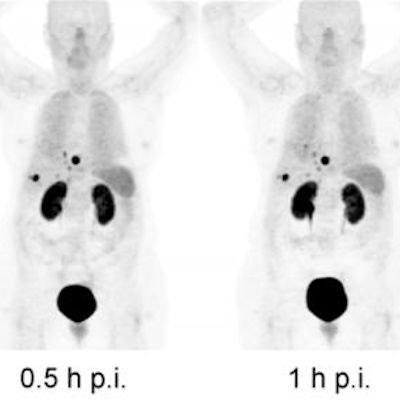
 PET images with Ga-68 OPS202 show that tumor and renal uptake persist over time while organs such as the spleen, lungs, and liver, along with the urinary excretion, are progressively washed out. The highest tumor contrast was found one hour after injection. Images courtesy of JNM.
"Even though the effective dose of Ga-68 OPS202 is comparable to other Ga-68-labeled somatostatin analogs, there are striking differences concerning its biodistribution and organ doses such as liver, gastrointestinal tract, pancreas, lung, and spleen," said study co-author Dr. Damian Wild, PhD, from University Hospital Basel in Switzerland, in a statement from the Society of Nuclear Medicine and Molecular Imaging (SNMMI).
In addition, Ga-68 OPS202 was well-tolerated, with no safety concerns among the 12 subjects.
"Ga-68 OPS202 could be a favorable alternative to the current radiolabeled somatostatin agonists in use in the clinic for PET/CT imaging of neuroendocrine tumor patients," Wild said. "Due to their enhanced binding properties, radiolabeled sst antagonists may open a new avenue for PET imaging and targeted radionuclide therapy in non-neuroendocrine tumor indications."
The findings warrant further evaluation of Ga-68 OPS202 for PET/CT imaging of NETs and other sst2 receptor-expressing tumors, the authors noted.