When I was a wee girl, my mother used to brag about being the only doctor in the chest division at the Hospital of the University of Pennsylvania to be able to really see chest x-rays in stereoscopic vision. I tried, years later as a student at Duke University, but alas I did not have her skill. My brain could not fuse the two images, and I could never place a nodule in space.
Did that make my mother a better imager than I, a radiologist? What is imaging anyway, and what defines an imager? Is an imager defined by specialty, by talent, by desire, or by something else? Is radiology itself an artificial construct, technology-based yet not useful?
No -- I don't think we can hang up our white coats yet!
Radiologists add value
Hot on the tails of attending this month's Imaging in 2020: The Future of Precision Medicine conference in Jackson Hole, WY, I am even more convinced that, yes, we do add value -- very important value. Imaging 2020 is an interdisciplinary gathering, designed to promote thought and conversation focused on state-of-the-art imaging, with a somewhat different theme each time.
And that is what led me to the question: What is imaging indeed?
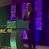
The most exciting talk of the meeting, at least by a consensus of radiologist attendees, was on Sunday evening, by Dr. Sanjay Jain from Johns Hopkins University.1 The topic was noninvasive ways to detect infection through bacteria-specific imaging tracers combined with CT imaging. Wow!
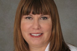
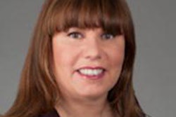
 Dr. Mary Morrison Saltz.
The case he described was of a child with multiple-drug-resistant tuberculosis after a trip to India, and you could watch in almost real-time as the infection cleared and response to antibiotics visualized. What a breakthrough, bringing experimental techniques to clinical practice, and so many other applications spring to mind. Can you imagine imaging a diabetic foot: Is it infected, yes or no? Is there an abscess or merely inflammation? Which organism?
And yet the cancer imagers were worried about the radiation dose from CT, which one could imagine they might be. Yet they did not seem to grasp the very important clinical conundrum faced every day in the practice of radiology, and how such a technique could be lifesaving and would change the daily practice of medicine.
One person asked me, "Why try to find out if it is infected, since you will treat with antibiotics anyway? Don't the risks from the radiation make this test useless?" In life and death infections, imagine watching the infection melt away, confirming response to treatment? Such an advance would change outcomes on a daily basis.
All of the radiologists in the audience were so excited about this talk, yet perhaps the rest of the audience was less so, seeing risks, problems, and less potential to be lifesaving.
Yet we were all imagers at Imaging in 2020 -- not just the radiologists, but also the basic scientists, surgeons, pediatricians, pathologists, and engineers. Their imaging approaches and their understanding is different from those of radiologists, but they are also imagers. A surgeon's need to know how to optimize laser therapy for port wine stains in real-time in the operating room (OR) is addressed by laser speckle imaging, and this is imaging today!2 What about imaging an avulsed hand after surgical reanastomosis by a microvascular surgeon? Visualizing blood flow confirms a good outcome with maximal probability for success.
One cannot excise that which one cannot see, or at least not easily. Fluorescence is a hugely important tool to identify and characterize tumors both in experimental animals and in the OR every day. This too is imaging, and it's mission-critical to surgical success. Yet this imaging is not radiology either.
We radiologists, the quintessential imagers, are not usually there in the OR working side by side with our surgeon colleagues. Are the surgeons indeed imagers too?
A lymphangiogram was a precursor to fluorescence, outlining the invisible and allowing us to see within the otherwise invisible lymph node. Perhaps this should remind us that the technologies of today will be replaced with new methods, but the quest to detect and understand the unseen has not really changed, although the methods have improved, as has the understanding.
Where do radiology, PET, and MRI meet chemistry, physiology, and function? PET/MRI is this crossroad, and one that is a new frontier in imaging. Understanding and targeting specific markers, and implementing them at once, allows us a window into where form meets function. Here we radiologists must partner with other clinicians, chemists, and biologists to design and execute studies leading us to increased understanding of pathophysiology. This can help us unlock answers to diseases such as cardiac sarcoidosis and epilepsy, where a functional and anatomic roadmap can teach us much about the underlying processes leading to the manifestation of diseases.
A whole new world of imaging
The keynote address at the Imaging 2020 meeting was by William Lange, a research specialist and director of the Advanced Imaging and Visualization Laboratory at Woods Hole Oceanographic Institution. He discussed imaging of the deep sea, specifically the use of extraordinary hardware and software to image its creatures, sea wrecks, and the ocean floor, giving us great insight into the ocean, its biodiversity, and what brings our mighty vessels to the floor of the sea. Each year 2,000 seafarers are lost to the waves, and discovering how and why is done through imaging.
Lange, too, uses image analysis and image feature extraction, just as we do in radiomics and pathomics -- the same imaging, but on a completely different scale, and yet to the same end of better understanding nature, where it goes astray, why, and how.
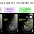
Imaging 2020 opened my eyes to a whole new world of imaging. We radiologists are part of a community of imagers, going well beyond what we may think of as radiology. Yet as radiologists we have a unique 360° view that is rare in medicine. It can be hard to quantify our abilities: Maybe sometimes we can just "smell cancer"3 or find a phone because we search with "radiology vision."
So, after all, perhaps my mother was indeed a better imager than I -- or at least we belong to the same club.
References
- Ordonez A, Weinstein E, Bambarger L, et al. A systematic approach for developing bacteria-specific imaging tracers. Journal of Nuclear Medicine. September 15, 2016.
- Huang Y, Ringold T, Nelson J, Choi B. Noninvasive blood flow imaging for real-time feedback during laser therapy of port wine stain birthmarks. Lasers in Surgery and Medicine. 2008;40(3):167-173.
- Evans K, Haygood T, Cooper J, Culpan A, Wolfe J. A half-second glimpse often lets radiologists identify breast cancer cases even when viewing the mammogram of the opposite breast. PNAS. 2016;113(37):10292-10297.
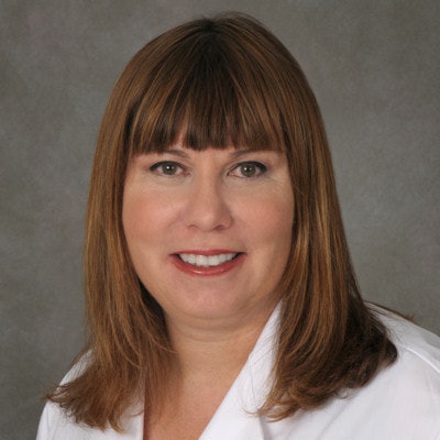
Dr. Mary Morrison Saltz is a board-certified diagnostic radiologist. She is currently chief medical information officer for the Stony Brook Cancer Center, a member of the Stony Brook department of radiology, an adjunct member of the department of biomedical informatics, and chief clinical integration officer for community practice initiatives at Stony Brook Medicine.
She has also served as chief medical officer for Hospital Radiology Partners and as radiology chair and service chief at hospitals in Florida, Ohio, and Georgia. Dr. Saltz is a member of the American College of Physician Executives, the American College of Radiology, and RSNA, and she sits on the Citizens Advisory Council of the Duke Cancer Institute.
A hands-on leader, Dr. Saltz's expertise is working hand-in-hand with hospital administration to guide radiology teams to success. Dr. Saltz has led quality assurance programs in Florida and Ohio, and she served as chief quality officer for community practice initiatives at Emory Healthcare. She also has more than 20 years of private-practice experience.
She is a graduate of McGill University, with a Bachelor of Science in Human Genetics, and Duke University, where she obtained her Doctor of Medicine. Her postgraduate education included a residency at Boston University, where she served as chief resident, and a fellowship in interventional abdominal radiology at Massachusetts General Hospital.
The comments and observations expressed herein do not necessarily reflect the opinions of AuntMinnie.com.
