
Top Story

Latest News


Sponsored
My BreastAI Suite by GE HealthCare
April 2, 2024

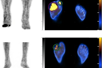
Cases of the Week
Check out our Cases of the Week!


More from AuntMinnie
Next Generation Radiology Workflow Tools
April 24, 2024

Voiant and Thirona enter commercial partnership
April 24, 2024

Spectral AI names CEO of Spectral IP, plans spin-off
April 24, 2024


