Ever wonder what man's best friend is really thinking? Georgia researchers are perhaps a step closer to the answer: Using functional MRI (fMRI) for the first time in unrestrained, unsedated dogs, they found that a hand signal associated with reward led to activation in a specific part of the dog's brain.
The fMRI scans showed significant activity in the caudate, a brain region linked to reward prediction, when two dogs were given a reward hand signal, but there was no such activity with a no-reward signal, the group found. The results were published May 11 in PLoS One.
The idea of training a dog to lie still for the duration of a scan -- as opposed to using restraints -- was sparked by military animals, said lead author Dr. Gregory Berns, PhD, director of the Center for Neuropolicy at Emory University in Atlanta. "I realized that if dogs can be trained to jump out of helicopters and airplanes, we could certainly train them to go into an fMRI to see what they're thinking."
 |
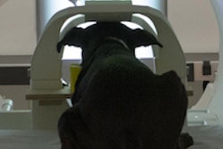
| One of the study participants, Callie, in front of the scanner. Image courtesy of Emory University. |
A new take on 'lie down'
"To our knowledge, this is the first demonstration of either MRI or fMRI in completely awake, unrestrained dogs," wrote Berns and co-authors Andrew Brooks, a graduate student at Emory, and Mark Spivak, a professional dog trainer. Cognition could not be studied if the animals were sedated, and the group wanted to avoid the immobilization techniques used in other animal studies.
 |
| Callie practicing in a replicated head coil. Image courtsey of Dr. Gregory Berns, PhD, and PLoS One. |
Replicas of the scanner were built for the dogs' homes or training areas, and after gradual acclimation to the replicas, the dogs were eventually transitioned to the actual scanner (3-tesla Magnetom Trio, Siemens Healthcare).
During the scanning session, the dog's handler faced the dog at the head of the scanner and gave hand signals associated with a food reward: The left hand up indicated the dog would receive a hot dog reward, whereas both hands pointing toward each other horizontally meant no reward, the group wrote.
The dogs had been previously trained on these signals, and the handler maintained each signal for approximately 10 seconds. After each reward trial, the handler would reach in to give the dog the reward.
Protocol for the trainees
The researchers obtained both functional and structural MRI scans of each dog. For the functional scans, they used single-shot echo-planar imaging, acquiring volumes of 28 sequential 3-mm slices with a 10% gap, the authors wrote.
 |
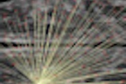
| Handler giving the reward hand signal. Image courtsey of Dr. Gregory Berns, PhD, and PLoS One. |
The researchers processed the data using Analysis of Functional Neuroimages (AFNI) software and corrected for motion. While the dogs did move between the trials (for example, to eat the food treat), the group found that they generally returned to the same position.
"Although the intertrial movements were large compared to humans, once set in the chin rest, the dogs were comparable, if not better, than humans," they wrote.
Caudate activation
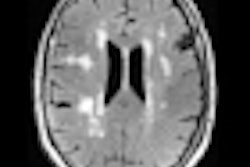
The fMRI scans showed significant activation in the right caudate in both dogs in response to the reward signal versus the no-reward signal, Berns and colleagues concluded.
"Although the statistical significance of the caudate cluster was modest in each subject individually (p < 0.01 in Callie, and p < 0.001 in McKenzie), the observation of the same location in the same condition in both dogs, and in the hypothesized region, strongly suggests that these were not spurious findings," they wrote.
 |
| In the above MR images, more activity is seen in the caudate with the reward signal compared to the no-reward signal. The image below represents both dogs, with the images co-registered and overlaid on Callie's structural scan. Images courtsey of Dr. Gregory Berns, PhD, and PLoS One. |
 |
"These results indicate that dogs pay very close attention to human signals, and these signals may have a direct line to the dog's reward system," Berns said.
Future research might investigate the effects of using an inanimate signal, such as a light, versus using the dog's handler, the authors wrote. This could help clarify whether the dog's reaction is solely based on the anticipated food reward, or whether the dog is also reacting to a possible social reward from the handler.
The fact that the dogs were able to remain motionless for up to 24 seconds -- willingly and using only positive reinforcement -- opens up many possibilities for future studies, according to the group.
"How do dogs distinguish humans, and is it by vision or smell?" they asked. "Is human language processed as arbitrary sounds, or do dogs have neural structures that respond in a deeper manner to language? What is the difference between how dogs represent humans and other dogs or animals? The questions are endless."