A group of German and U.S. researchers and engineers is showing early success with a prototype hybrid PET/MRI system, which features a full-ring PET insert installed inside a 7-tesla MR scanner.
One goal of the collaboration among the University of Tübingen in Germany; Bruker BioSpin MRI of Ettingen, Germany; and Siemens Preclinical Solutions of Knoxville, TN, is to expand functional imaging and merge function with anatomy into one device.
"MRI gives us a higher resolution and soft-tissue contrast. With PET, we get high sensitivity and a variety of tracers," said Hans Wehrl from the University of Tübingen's Laboratory for Preclinical Imaging and Imaging Technology. "We don't get additional radiation dose in contrast to PET/CT, and we save on anesthesia time."
Wehrl presented the results of the group's study to investigate the performance of a hybrid PET/MRI small animal imaging system at the 2007 joint conference of the Academy of Molecular Imaging (AMI) and the Society of Molecular Imaging (SMI) in Providence, RI.
"Now that we can, for the first time, image in the functional process with MRI and PET simultaneously, we can get the same compounding parameters," he said. "We can look at the same parameters, such as perfusion MRI and PET, or we can look at different functional process, such as perfusion in PET and BOLD imaging in MRI."
Conflicting technologies
The main issue in combining the two modalities into one hybrid system is how the technologies will react to each other in such close proximity. To determine the effect, the team placed a full-ring PET insert inside the MRI scanner. Phantom tests were performed to assess potential interference with the PET insert inside the MRI system. Signal-to-noise ratio and image homogeneity were evaluated for turbo spin-echo (TSE) and gradient-echo (GRE) sequences.
The configuration offered a combined field-of-view of 90 mm in the axial direction and 8 mm in the transaxial direction. MR spectroscopy was performed for different voxel volumes and positions inside the PET/MRI field-of-view. BOLD imaging stability also was evaluated according to Function Biomedical Informatics Research Network (FBIRN) guidelines.
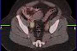
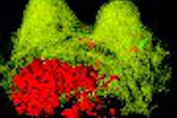
Simultaneous in vivo data of a mouse brain were collected with MRI and PET using the dopamine transporter ligand C-11 methylphenidate. A mouse brain was studied with PET/MR after injecting simultaneously a gadolinium MR contrast agent and O-15 water as a PET tracer.
Using various different imaging sequences, the group found the same level of signal-to-noise ratio regardless if the PET insert was inside the MRI system or not (without/with the PET insert: TSE 446/440, GRE 66/64). The same results held true for image homogeneity with little difference in the location of the PET insert (without/with the PET insert: TSE: 95/97, GRE: 87/86).
In MRI spectroscopy studies on a phantom, spectral quality remained high. "This is an indication that we don't have a lot of anti-current defects caused by the PET insert," Wehrl said.
Image distortion
The placement of the PET insert inside the MRI scanner and changing gradient strengths to acquire certain MRI images may reduce the efficiency of the gradient strengths and result in image distortions, Wehrl noted. One way to inspect and correct for the problem is to take the MRI images with and without the PET insert and perform image subtraction.
The team successfully used image subtraction for different sequences to correct for image distortion. "Even in the case of planar imaging, which is a very sensitive image sequence, we only see very small variations on the order of 1 pixel size," Wehrl said. "We think these differences are caused by the fact that we have to reposition the system and the phantom and not adjust the PET insert itself."
Functional imaging is possible in a configuration that features a PET insert inside an MRI scanner, the group concluded. "On the PET side, we get the same performance inside and outside of the MR system in terms of sensitivity and spatial resolution," Wehrl said. "We can acquire very high-quality PET and MRI images, which do not have to be compromised by combining these two modalities."
By Wayne Forrest
AuntMinnie.com staff writer
October 10, 2007
Related Reading
Zecotek, UW collaborate on PET/MRI, July 27, 2007
PET/MRI system an ISMRM highlight for Siemens, May 22, 2007
First PET/MRI brain images debut at SNM 2007, June 15, 2006
Copyright © 2007 AuntMinnie.com