When the information systems group at the Medical University of South Carolina (MUSC) last year brought online a 20-TB storage area network (SAN), the team figured the Charleston medical school had enough archive storage capacity to handle patient images for the next three years.
Healthcare providers and radiologists then added more multislice CT, MR, computed radiography (CR), digital mammography, and a host of other modalities' images to the PACS. Suddenly, the amount of free space on the SAN began to dwindle quickly.
"At the rate we are currently using it, we now have less than two years of online storage available on those 20 terabytes," said Eugene Mah, medical physicist at MUSC. "A couple of years ago, we could have kept 24 months of data online on less than 7 terabytes. Now we need at least 25 terabytes."
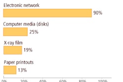
What happened to all that newly installed capacity? The four biggest storage-gobbling culprits are CT, CR, MR, and digital mammography, which cumulatively consume approximately 98% of MUSC's storage requirements. "These four modalities are the biggest contributors for future planning or expanding a PACS or archive," Mah told attendees at the recent 2007 Society for Imaging Informatics in Medicine (SIIM) meeting in Providence, RI.
Good old days
Before the voluminous images of multislice CT and digital mammography came along, it was a relatively easy task to estimate future archive storage needs. Estimate the storage size for CR images, add 50% for the other modalities, and the forecast was done.
To get a better handle on the situation at MUSC, Mah and his colleagues reviewed the past 11 years' worth of data in the medical school's PACS, pouring over CT, CR, MR, digital mammography, PET/CT, ultrasound, and nuclear medicine images to determine how much archive storage space each modality required. "Based on the image file sizes, we were able to compute how many gigabytes per month we were archiving for each modality," Mah said.
Every image at MUSC is archived to its PACS, because that is the model the radiologists want. "It provides better data integrity and everything is validated to the RIS," Mah noted. "It allows the radiologist to retrospectively reconstruct any dataset or prior comparisons." The downside, he added, is increased storage consumption and storage costs.
Constant upgrades
In this decade alone, MUSC has made a multitude of advances to its imaging technologies. In 2000 and 2001, all of its CT scanners were upgraded from single-slice to dual- and four-slice CT systems. In 2003, the dual- and four-slice CT scanners were replaced with 16-slice CT units. By 2005, one 16-slice CT machine was replaced with a 64-slice model, and last year one 16-slice CT scanner was replaced with a dual-source CT system.
In addition, MUSC converted four mammography units to digital capabilities in 2005. Five years ago, the institution added a 3-tesla MRI system and installed an open MRI magnet two years ago. Last year, the medical facility upgraded its PET scanner with 16-slice CT technology.
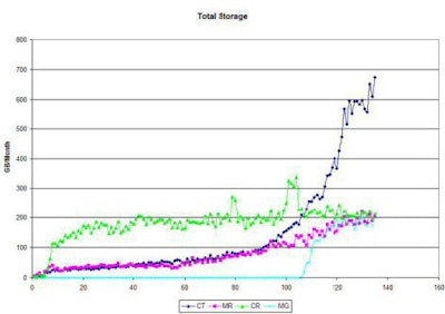
At one time, CR was the dominant consumer of archive storage pace. The MUSC study found that much of the modality's growth in recent years has resulted from MUSC adding new service locations, rather than increased volume. "Over the past 11 years, we have seen a 50% increase in the number of CR studies stored to the archive," Mah said. "The total image volume, however, has been fairly stable. As a result, the total storage, in terms of gigabytes, has been fairly stable as well."
MUSC's CT scanners continue to output more images to the archive. Radiologists are performing more 3D reconstructions and multiplanar reformats (MPRs), which means studies routinely can contain as many as 10,000 images.
Not surprisingly, the rapid, exponential spike in the number of CT images archived each month corresponds with MUSC's upgrade from single-, dual-, and four-slice CT scanners to multidetector CT systems. In fact, the number of CT images going to the PACS has doubled over the past 14 months, while the average number of images per study has doubled within the last 22 months.
CT dominates
Digital mammography, meanwhile, had "a very unexpectedly large effect on storage consumption," Mah recalled. Even though MUSC has only four digital mammography units, they combine to occupy approximately 17% of the available storage space.
 |
| CR's impact on storage capacity has remained relatively stable, while multidetector CT and digital mammography (MG) increasingly occupy more storage space. Chart courtesy of Eugene Mah, MUSC. |
"With the installation of the multidetector CT scanners, CT is becoming the dominant modality for images," Mah noted. "We see similar behavior when it comes to storage factors. At the present time, CT makes up about 50% of our full storage usage. CR, digital mammo, and MR are each using about 15% of storage."
The MUSC study also reviewed other modalities -- such as ultrasound, angiography, digital fluoroscopy, and nuclear medicine -- and found that they accounted for only about 10% of the total storage. The analysis did not prompt MUSC to significantly change its storage distribution, although it did increase the contribution of ultrasound.
 |
|||
| Image File Sizes |
|||
| Modality | Matrix Size | File Size (MB) | |
|
|||
| CT | 512² | 0.5 | |
|
|||
| MR | 512² | 0.5 | |
|
|||
| CR | 2048 x 2494 | 10 | |
|
|||
| Nuclear medicine | 256² | 0.128 | |
|
|||
| Digital mammography | 4096 x 3328 | 26.6 | |
|
|||
| PET/CT | 128² | 0.032 | |
|
|||
| US | 640 x 480 | 0.3 | |
|
|||
| By estimating the file sizes for each modality, MUSC can forecast its future storage capacity needs. Data courtesy of Eugene Mah, MUSC. | |||
Storage requirements for each MUSC modality are computed using estimated file sizes. "We can take this data and use it to project how many months of data we can store online," Mah said, "and we can project how much storage space we need to maintain a certain number of months of data online."
To forecast future archive storage needs, Mah recommends that healthcare facilities gather monthly storage data from the last 12 months, and collect the number of images per month for CT, CR, MR, and digital mammography. Then, estimate the total storage per month based on image file sizes and examine the internal trends. That model should forecast future storage needs, based on the growth of a facility's radiology services.
By Wayne Forrest
AuntMinnie.com staff writer
June 18, 2007
Related Reading
Software facilitates inclusion of nonradiological images into PACS, January 16, 2006
Choose image archiving solutions before data volume grows too large, January 5, 2006
Digital library can ease transition to digital mammo, October 26, 2005
FDA ruling opens door to new PACS storage options, July 24, 2003
Copyright © 2007 AuntMinnie.com