Physicians and radiologists may have another hybrid imaging technology to delve deeper into the burgeoning realm of molecular imaging and investigate neurological studies, as well as certain forms of cancer, stroke, and stem cell therapy.
The technological development comes from researchers at the University of Tübingen in Germany, the University of Tennessee in Knoxville, and Erlangen, Germany-based Siemens Medical Solutions. The collaborative team unveiled the world's first human brain images from a work-in-progress PET/MRI system at the 2007 SNM meeting last week in Washington, DC.
Bernd Pichler, Ph.D., head of the Laboratory for Preclinical Imaging and Imaging Technology at the University of Tübingen, told attendees that the PET/MRI system could help researchers better understand the pathologies and progression of neurological disorders, such as Alzheimer's and Parkinson's diseases, epilepsy, depression, and schizophrenia.
The PET/MRI system is located at Siemens' factory in Knoxville. A patient who is referred for a PET/CT scan also may receive a PET/MRI brain scan at the Siemens facility, if he or she signs a consent form. The research team began scanning patients late last year.
"There has been a period of rebuilding and redesigning the PET detectors, so we haven't done any recently, but we're hoping that will pick up again," said David Townsend, Ph.D., director of the Molecular Imaging and Translational Research Program at the University of Tennessee Graduate School of Medicine.
 |
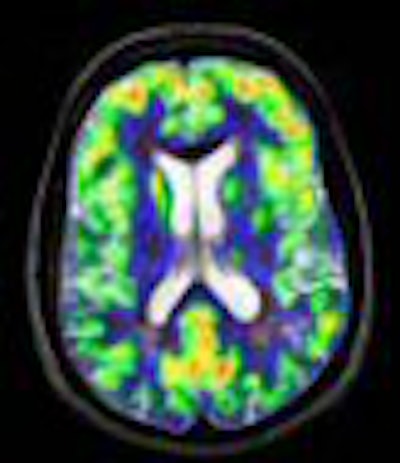
| Brain PET and Siemens' Magnetom Trio MRI feature a removable PET head insert. Image courtesy of Siemens Medical Solutions. |
Scanner design
The prototype dedicated brain PET scanner uses next-generation avalanche photodiode detector (APD) technology to render the PET scanner impervious to magnetic fields and, in turn, not interfere with the imaging capabilities of MRI. The PET scanner is integrated into a standard Siemens 3-tesla Magnetom Trio MRI unit. The combined system has a spatial resolution of 3 mm, axial field-of-view of 19 cm, and transaxial field-of-view of 30 cm.
One key to the clinical success of the PET/MRI system is its configuration. Simply attaching a PET scanner to a standard MRI system -- similar to a PET/CT design -- would produce a sequential scan, rather than a simultaneous scan. The result would mean a longer scan time for the patient and no temporal correlated data between PET and MRI, Pichler said.
"The ultimate goal would be to completely integrate the PET scanner into the MRI scanner," he added in his SNM presentation, "to have this process in combined imaging, temporal correlated data, and a short scan time."
 |
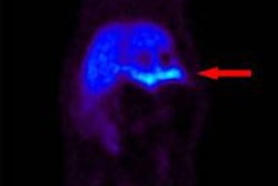
| FLAIR, ADC, T1 MPRAGE, and T2 TSE were applied during the same PET acquisition. PET shows absence of FDG signal from pathological periventricular regions. Images courtesy of Bernd Pichler, Ph.D., and colleagues at the University of Tübingen and David Townsend, Ph.D., and colleagues the University of Tennessee. |
The team also has been working to rectify the issue of attenuation correction, which for an MRI image is much more challenging than on a CT image, Pichler said. Using animal scans, researchers believe they have more than adequately matched PET and MRI scans. "It is definitely possible to do attenuation correction based on an MRI image and get a quantitative PET/MR image modality," he added.
The study noted that neither the MRI nor the PET images were degraded by the synchronous data acquisition.
Pichler said the benefits of the PET/MRI system would be "simultaneous measurement with perfect anatomical matching and temporal correlation of PET/MRI data," adding that the combined imaging modalities show "excellent soft-tissue contrast," and gating and motion correction can be possible through MRI.
Townsend added that simultaneous molecular and spectroscopy or anatomical imaging with MR have potential applications. "We'll find out whether it remains a strong research application," he said. "There are clinical applications, so radiology departments would buy a molecular imaging ring, a PET insert, and insert it into their MR scanner."
 |
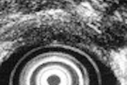
| Comparative brain images with diffusion echoplanar imaging sequence applied during PET acquisition. Images courtesy of Bernd Pichler, Ph.D., and colleagues at the University of Tübingen and David Townsend, Ph.D., and colleagues at the University of Tennessee. |
Townsend was influential in the development of PET/CT during his tenure at the University of Pittsburgh in Pennsylvania in the late 1990s and earlier this decade. The first PET/CT images were taken in 1998; the first commercial PET/CT systems hit the market in 2001.
In comparing the two hybrid technologies, Townsend said they "had a very different initiation of growth." "The PET/CT was built initially when we built that prototype as a scientific project where we felt that offering clinical anatomy and clinical functional imaging made a lot of sense," he added. "MR is a much more challenging environment in which to combine with molecular imaging."
PET/MRI has been "a commercial development," Townsend continued, "because very few people knew about it outside the factory. It wasn't really something that the physicians demanded. Now we have the capability of PET/MRI, which has been a technological breakthrough in getting these detectors to work, we are going to have to see what sort of demand comes from the medical side."
By combining PET and MRI, the researchers said clinicians also hope to make a more definitive determination of cognitive impairment and atrophy, assessing those conditions with the advent of new neurological biomarkers. In addition, the hybrid modality may help doctors locate salvageable brain tissue in stroke patients.
PET can differentiate mild cognitive impairment from early-stage Alzheimer's, but the technology cannot determine reduced brain volume caused by atrophy.
With MRI strong in areas such as the central nervous system, brain, and joints, the ultimate goal of the collaboration is to develop a whole-body PET/MRI system.
By Wayne Forrest
AuntMinnie.com staff writer
June 15, 2007
Related Reading
Molecular imaging, nuclear medicine share spotlight at SNM 2007, June 4, 2007
Siemens unveils HD-PET, June 4, 2007
PET/MRI system an ISMRM highlight for Siemens, May 22, 2007
Copyright © 2007 AuntMinnie.com