Japanese researchers using MRI have found that longer a person has diabetes, the higher their risk of gray-matter atrophy, according to a study published May 2 in Diabetes Care.
And since gray-matter atrophy is associated with diseases such as Alzheimer's, the findings shed light on the long-term negative effects of diabetes, wrote a team led by Dr. Naoki Hirabayashi of Kyushu University in Fukuoka, Japan.
"[Our] study suggests that the longer duration of diabetes and a midlife onset of [it] are significant risk factors for gray matter atrophy in various brain regions that are likely to play an important role in cognitive function," the group noted.
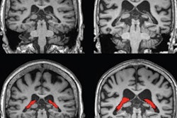
Epidemiological studies have suggested that diabetes is associated with risk of developing dementia, but this research has only explored links between diabetes and total brain and hippocampal volume, the authors explained. They sought to refine the investigation of a connection between diabetes and brain volume -- and thus dementia risk -- using voxel-based morphometry methods with brain MRI to measure specific regions. Voxel-based morphometry can identify subtle structural changes in the brain that may be connected to neurological dysfunction.
The team used data from the Hisayama Study, a population-based observational effort to assess cardiovascular disease and dementia in Japan that launched in 1961. For the current study, Hirabayashi's team included information from 1,189 Japanese people aged 65 and older who underwent brain MRI exams in 2012. Of these, 272 (23%) had diabetes.
The researchers measured participants' regional gray matter volumes and intracranial volume by applying voxel-based morphometry techniques to the MRI data. They then analyzed any associations between diabetes and parameters of the regional gray matter volumes and intracranial volumes using an analysis of covariance and regression (ANCOVA) model and evaluated regional gray matter atrophy with the voxel-based morphometry method.
The ANCOVA analysis showed that those study participants with long-term diabetes (that is, circa a 1988 health examination conducted for the Hisayama study) had statistically significant lower mean values of gray-matter and intracranial volumes compared with healthy counterparts in the frontal lobe, the temporal lobe, the insula, in deep gray-matter structures, and in the cerebellum.
The voxel-based morphometry analysis showed that diabetes was associated with gray-matter atrophy in the following brain areas:
- Bilateral superior temporal gyri
- Right middle temporal gyrus
- Left inferior temporal gyrus
- Right middle frontal gyrus
- Bilateral thalami
- Right caudate
- Right cerebellum
The team also found that gray-matter and intracranial volumes were lower in those study participants with long-term elevated two-hour postload (after a meal) glucose levels, a finding that could be key to tailoring preventive dementia care for diabetics.
"The significant association between elevated two-hour postload glucose levels and gray-matter atrophy in various brain regions may suggest that careful control of postprandial plasma glucose levels is important to prevent gray-matter atrophy and the subsequent development of dementia in individuals with diabetes," the authors concluded.