Researchers have developed a low-dose, multicontrast chest x-ray system that offers the potential to improve the clinical diagnosis and screening of lung diseases, according to a November 29 presentation at RSNA 2021 in Chicago.
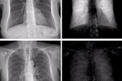
Researchers from the University of Wisconsin-Madison designed and built a chest x-ray prototype that acquires conventional absorption contrast, differential phase contrast, and dark-field x-ray contrast images in a single shot and breath hold.
"This technique can provide three complementary images which can be jointly used to enhance the imaging functionality of chest radiography," said medical physicist Ran Zhang, PhD.
While chest x-ray is the most frequently performed diagnostic radiography imaging test, it is limited in certain cases by overlapping anatomy and is less sensitive imaging low attenuating lesions. Pulmonary lesions are often blocked by overlapping ribs, for instance, and they can be difficult to detect due to their relatively low contrast compared with surrounding tissue.
To overcome these limitations, Zhang and colleagues built a system that extracts differential phase and dark-field contrast information as well as conventional attenuation contrast images. The system employs a standard radiography tube and flat-panel detector with the addition of three grated interferometers between them mounted on a separate gantry. This reduces vibrations, Zhang said.
"The grating interferometer generates interference fringes that carry the attenuation phase and dark-field information of the imaging object," Zhang explained.
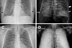
 Image courtesy of Ran Zhang, PhD.
During data acquisition, subjects are scanned in pulse mode (30 pulses per second) while the table moves along the superior-inferior (SI) direction. The detector readout is synchronized with the pulses. A slit-scan design allows scanning of the entire chest with a lateral coverage area of 32 centimeters along the SI direction with a scan speed of 7 cm per second, Zhang said.
"That means the whole lungs can be covered in less than 5 seconds or in a single breath hold," he said.
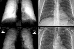
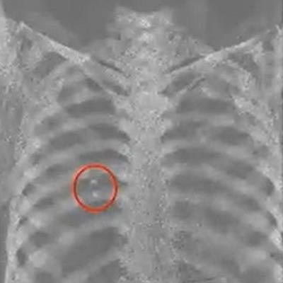
Initial tests were performed using an anthropomorphic chest phantom. To simulate potential lung nodules, a container containing microbubble powder and calcium hydroxyapatite was attached to the chest phantom.
The estimated air kerma and effective dose were 53 uGy and 9.2 uSv, which is well below the effective dose for a typical chest x-ray (~20 uSv), Zhang said. The quality of the absorption contrast image matched that of conventional chest x-rays. In addition, the dark-field image revealed the microbubble and the calcium hydroxyapatite, which was invisible in the absorption image due to anatomical noise, Zhang said.
"Preliminary phantom and animal imaging results show promising image quality and contrast levels," he said.
Zhang noted the microbubbles used to simulate lesions are powders that simulate microstructures that generate strong contrast, and whether they accurately simulate lung lesions is a question that remains to be explored.
Future work will focus on improving the signal-to-noise ratio of the dark-field image and reducing image artifacts, he concluded.