Routine chest CT scans offer body composition information that could help clinicians better evaluate the mortality risk of patients with chronic obstructive pulmonary disease (COPD), according to a study published April 6 in Radiology.
This additional data could translate to mitigated risk due to earlier intervention, a team led by Dr. Farhad Pishgar of Johns Hopkins University School of Medicine in Baltimore said in a statement released by the journal.
"In theory, CT-derived body composition assessments would provide an opportunity for earlier interventions in patients who face a higher risk of adverse health events," the group said.
Chest CT has long been used to assess lung health in people with COPD, but the exam could also offer a way to evaluate obesity and sarcopenia through soft-tissue biomarkers. Pishgar's group used chest CT data to investigate links between imaging-based soft-tissue markers and all-cause mortality in patients with COPD.
The study included information from 2,994 patients enrolled in the Multi-Ethnic Study of Atherosclerosis (MESA) who underwent routine chest CT between 2010 and 2012; of these, 265 people had COPD. The group extracted mortality data from the National Death Index, finding that 49 (18%) of the COPD patients died of the condition over the follow-up period.
The team found that fat levels of the COPD patients affected mortality rates, but this effect depended on what type of fat: Those with greater intermuscular fat had higher all-cause mortality rates, while higher levels of subcutaneous fat tissue were associated with lower risk of all-cause mortality.
| Association of imaging-derived markers with all-cause mortality among patients with COPD | ||
| Characteristic | Unadjusted hazard ratio | Adjusted hazard ratio* |
| Subcutaneous adipose tissue index | 0.58 | 0.22 |
| Intermuscular adipose tissue index | 1.26 | 1.39 |
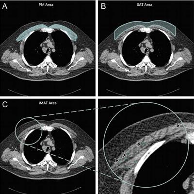
 Axial chest CT examination in a 54-year-old participant. (A) On the axial noncontrast chest CT image, the pectoralis muscle (PM) area was segmented and measured in the section above the aortic arch. (B) The subcutaneous adipose tissue (SAT) area as the area between the pectoralis muscle and the skin surface on the same section was also measured and the attenuation of pixels in the subcutaneous adipose tissue area was used to determine the individualized threshold for the intermuscular adipose tissue. (C) The intermuscular adipose tissue within the pectoralis muscle was segmented as the areas with Hounsfield units below this threshold for the intermuscular adipose tissue (IMAT) (arrowheads). Images and caption courtesy of the RSNA.
"Compared with subcutaneous adipose tissue quantification, intermuscular adipose tissue may be a better marker for predicting all-cause mortality in patients with COPD ... [in part because] the intermuscular adipose tissue index is an indicator of other underlying comorbidities (e.g., diabetes and hypertension) and may predict the all-cause mortality better," the authors wrote.
The study suggests that soft-tissue markers identified on chest CT offer a reliable way to estimate mortality risk in COPD patients, wrote Dr. Nicola Sverzellati, PhD, and colleague Dr. Filippo Cademartiri, PhD, both of the University of Parma in Italy, in an accompanying commentary.
"Chest CT has the potential to become a powerful tool in the quest for personalized medicine in COPD," Sverzellati and Cademartiri wrote. "Whether it is incorporated into routine assessment for patients with COPD will ultimately depend on our ability to demonstrate that this information changes treatment and improves outcomes."