Are gadolinium-based contrast agents (GBCAs) really needed for breast MRI? Not if the technique of diffusion-tensor imaging (DTI) is used, according to a study published August 6 in the American Journal of Roentgenology.
Canadian researchers found that the combination of DTI with breast MRI shows equivalent accuracy for cancer diagnosis whether or not GBCAs are used -- results that would reduce women's exposure to gadolinium agents, the safety of which has been controversial.
In fact, not administering contrast makes DTI easier to use, said a team led by Dr. Anabel Scaranelo of Princess Margaret Cancer Centre in Toronto in a statement released by the American Roentgen Ray Society (ARRS).
"Contrast-enhanced DTI parameters are less amenable to standardization because their values depend on the type and dose of contrast agent and on variations in the contrast agent dynamics among the various types of breast cancers," Scaranelo and colleagues said.
The effects of GBCAs on DTI's performance haven't been clarified, according to the investigators. To address the knowledge gap, Scaranelo's team conducted a study that included 26 women with BI-RADS category 0, 4, 5, or 6 breast lesions on mammography who underwent two 3-tesla breast MRI exams, using the same DTI sequence before and after contrast.
The investigators used DTI software to create parametric DTI maps of directional diffusion coefficients (DDC), mean diffusivity, and maximal anisotropy index (measured by eigenvalues λ1 to λ3) of the lesions and of normal tissue.
Of the 26 women, 58% had cancer and 42% had benign or normal results on MRI. All of the cancers were identified in the DDC maps both before and after the women were administered contrast; the mean cancer size detected was 15.3 mm before contrast administration and 17.3 mm after contrast, a difference that was not statistically different (p = 0.35), the group found.
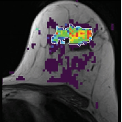
 61-year-old woman with grade 3 invasive ductal carcinoma of no special type and ductal carcinoma in situ mass lesion. Axial dynamic contrast-enhanced subtracted image (A) and directional diffusion coefficient λ1 parametric maps (B and C) overlaid on T1-weighted image of central slice with unenhanced (B) and contrast-enhanced (C) administration are shown. There is substantial contrast in λ1 map between normal tissue (purple, with λ1 ≥ 1.7 × 10⁻³ mm2/s) and cancer (green-yellow-blue, λ1 < 1.2 × 10⁻³ mm2/s). Lesion is manually delineated in B and C. Units in B and C are in mm2/s × 10–3. Images and caption courtesy of the American Roentgen Ray Society.
The study did show some change in imaging parameters after GBCA use: DDCs, mean diffusivity, and b0 signal intensity were lower in malignant lesions on GBCA imaging compared with imaging before GBCA use. Scaranelo's team also found that, with the exception of eigenvalue λ3, mean area under the receiver operating curve (AUC) values before and after the use of GBCA were not statistically significantly different across all parameters, ranging from 0.74 to 0.99 before contrast and 0.88 to 0.99 after contrast.
The study results confirm that using contrast for breast DTI-MRI isn't necessary. In fact, it may be better not to use it, the team concluded.
"Diagnostic accuracy using DTI was equivalent before and after GBCA administration, despite a change in the values of the DTI parameters," Scaranelo and colleagues wrote. "However, the limitations in standardization of contrast enhancement implies that unenhanced diffusion measurements should be preferred."