An imaging technique that combines F-18 FDG PET and MRI was able to pinpoint the origins of chronic pain in patients and help their caregivers develop better management plans, according to a Monday presentation at the Society of Nuclear Medicine and Molecular Imaging (SNMMI) 2020 virtual annual meeting.
The findings could offer relief to the millions of adults who live with chronic pain, according to a statement released by the SNMMI.
"Pain is the most common reason to seek medical attention, and those who suffer from it outnumber those who suffer from cancer, heart disease, and diabetes combined," the society said in the statement. "Chronic pain affects nearly 50 million adults in the United States and costs the nation's healthcare system as much as $635 billion in total expenses, including imaging and treatment costs."
Chronic pain is often caused by inflamed tissues that are highly metabolic and therefore good candidates for the glucose uptake generated by radiolabeled agents used in PET imaging, according to a team of researchers from Stanford University. The group sought to identify where pain originates in the body by combining anatomic information from MRI with molecular information from PET.
"In the past few decades, we have confirmed that anatomic-based imaging approaches, such as conventional MRI, are unhelpful in identifying chronic pain generators," said contributing study author Dr. Sandip Biswal in the SNMMI statement. "We know that [F-18] FDG PET has the ability to accurately evaluate increased glucose metabolism that arises from acute or chronic pain generators. As such, in our study we examined PET/MRI as a potential solution to determine the exact molecular underpinnings of one's pain."
The research included 65 patients with chronic pain who underwent whole-body PET/MRI. Biswal and colleagues used image analysis software to measure maximized standardized uptake values (SUVmax) on the F-18 FDG images; two radiologists assessed the images for correlations between SUVmax and pain sites.
In 58 of 65 patients (89%), the researchers found increased uptake of F-18 FDG in nerves and muscle at the site of pain, resulting in a slight change of management in 16 patients (such as an additional diagnostic test) and a significant change of management in 36 individuals (such as a new invasive procedure). The findings resulted in new management plans for 40 out of 65 patients (62%).
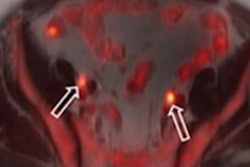
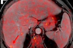
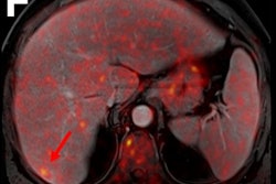
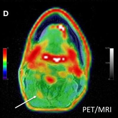
![Adult male with decades of right neck pain, discomfort, and tightening following birth injury. The patient had failed multiple standard therapeutic maneuvers before presenting for F-18 FDG PET/MRI. Images show abnormally elevated FDG uptake (white arrows; SUVmax = 1.2) observed in a linear pattern in the space in the posterolateral right neck, between the oblique capitis inferior and the semispinalis capitis muscles, where the greater occipital nerve resides. By comparison, the same region on the contralateral, asymptomatic side of the neck has an SUVmax = 0.7. This result encouraged a surgeon to explore the area. The surgeon ultimately found a collection of small arteries wrapped around the nerve in this location. The small arteries underwent lysis by the surgeon and the patient reported tremendous relief of symptoms. (A) Coronal thick slab MIP [maximum intensity projection] of F-18 FDG PET. (B) Axial LAVA [Dixon liver acquisition with volume acquisition] Flex MRI through the cervical spine. (C) Axial PET at the same slice as the axial MRI. (D) Fused axial PET/MRI. Image courtesy of Peter Cipriano of Stanford University; caption courtesy of SNMMI.](https://img.auntminnie.com/files/base/smg/all/image/2020/07/am.2020_07_14_17_31_5604_2020_07_13_SNMMI_PET_CT_chronic_20200714171056.png?auto=format%2Ccompress&dpr=2&fit=max&q=70&w=700) Adult male with decades of right neck pain, discomfort, and tightening following birth injury. The patient had failed multiple standard therapeutic maneuvers before presenting for F-18 FDG PET/MRI. Images show abnormally elevated FDG uptake (white arrows; SUVmax = 1.2) observed in a linear pattern in the space in the posterolateral right neck, between the oblique capitis inferior and the semispinalis capitis muscles, where the greater occipital nerve resides. By comparison, the same region on the contralateral, asymptomatic side of the neck has an SUVmax = 0.7. This result encouraged a surgeon to explore the area. The surgeon ultimately found a collection of small arteries wrapped around the nerve in this location. The small arteries underwent lysis by the surgeon and the patient reported tremendous relief of symptoms. (A) Coronal thick slab MIP [maximum intensity projection] of F-18 FDG PET. (B) Axial LAVA [Dixon liver acquisition with volume acquisition] Flex MRI through the cervical spine. (C) Axial PET at the same slice as the axial MRI. (D) Fused axial PET/MRI. Image courtesy of Peter Cipriano of Stanford University; caption courtesy of SNMMI.
Adult male with decades of right neck pain, discomfort, and tightening following birth injury. The patient had failed multiple standard therapeutic maneuvers before presenting for F-18 FDG PET/MRI. Images show abnormally elevated FDG uptake (white arrows; SUVmax = 1.2) observed in a linear pattern in the space in the posterolateral right neck, between the oblique capitis inferior and the semispinalis capitis muscles, where the greater occipital nerve resides. By comparison, the same region on the contralateral, asymptomatic side of the neck has an SUVmax = 0.7. This result encouraged a surgeon to explore the area. The surgeon ultimately found a collection of small arteries wrapped around the nerve in this location. The small arteries underwent lysis by the surgeon and the patient reported tremendous relief of symptoms. (A) Coronal thick slab MIP [maximum intensity projection] of F-18 FDG PET. (B) Axial LAVA [Dixon liver acquisition with volume acquisition] Flex MRI through the cervical spine. (C) Axial PET at the same slice as the axial MRI. (D) Fused axial PET/MRI. Image courtesy of Peter Cipriano of Stanford University; caption courtesy of SNMMI.The study suggests that this PET/MRI combination could be an effective way for those who experience chronic pain to get relief, Biswal said in the society statement.
"The results of this study show that better outcomes are possible for those suffering from chronic pain," he said. "This clinical molecular imaging approach is addressing a tremendous unmet clinical need, and I am hopeful that this work will lay the groundwork for the birth of a new subspecialty in nuclear medicine and radiology."