The common MRI technique called T1 mapping -- typically used for patients with heart disease -- could help clinicians evaluate the severity of neuroblastoma in children and the efficacy of treatment, according to results from an animal study published June 28 in Cancer Research.
The findings indicate that there could be an effective way to tailor treatments in children with neuroblastoma, suggested a team led by Dr. Yann Jamin from the Institute of Cancer Research in London, the U.K.
"T1 mapping scans could improve the use of precision medicine in children with neuroblastoma -- and potentially in cancer patients more widely -- by ensuring treatments are tailored for each patient and rapidly stopped when they are not working," the institute said in a statement.
T1 mapping indicates how water molecules intermingle at the cellular level and is often used to assess damage to the heart muscle, Jamin and colleagues wrote. But it appears to also be sensitive to MYCN, a protein linked to aggressive forms of neuroblastoma.
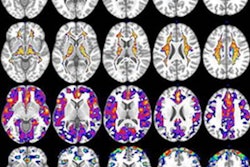
For their study, the investigators used artificial intelligence (AI) to map different cell populations of tumors in mice, then compared them with maps created using the T1 technique. They found an association between regions of the brain with high T1 values and more aggressive cancer cells, as well as a link between areas with low T1 values and benign tissue.
The team evaluated whether the T1 technique could assess how mice with neuroblastoma would respond to two drugs, alisertib and vistusertib, which target MYCN, a protein linked to aggressive forms of the disease. Jamin and colleagues found that the two drugs did stop tumor growth and decreased T1 values.
"In this study we demonstrate how T1 mapping is sensitive to the rich histological presentation of neuroblastoma, and can provide a sensitive biomarker of response to two clinically-relevant MYCN-targeted therapeutics in the Th-MYCN GEM model of neuroblastoma," the group wrote.
The study findings suggest a relatively easy way to better assess children with neuroblastoma, according to the group.
"Our findings show that an imaging technique readily available on most MRI scanners has the potential to pick out children with aggressive cancer and give us early signs of whether a treatment is working," Jamin said in the statement. "It is easy to perform and analyze T1 MRI scans, and they could be used to provide insights into many aspects of cancer biology -- and help doctors to design tailored treatments based on how aggressive a tumor appears to be."