An artificial intelligence (AI) model was able to predict a patient's risk for Alzheimer's disease by analyzing brain MRI exams and nonimaging factors such as age, gender, and cognitive test scores, according to research published online May 1 in Brain.
After training a deep-learning algorithm using data from the Alzheimer's Disease Neuroimaging Initiative (ADNI), a team of researchers from Boston University found that their model was highly accurate on three independent datasets. It also outperformed neurologists.
"This framework provides a clinically adaptable strategy for using routinely available imaging techniques such as MRI to generate nuanced neuroimaging signatures for Alzheimer's disease diagnosis, as well as a generalizable approach for linking deep learning to pathophysiological processes in human disease," wrote the researchers, led by Shangran Qiu, Prajakta Joshi, Matthew Miller, and Chonghua Xue.
Surmising that the clinical potential of deep learning is being attenuated by a lack of external validation and the use of opaque decision-making frameworks, the researchers sought to develop an explainable algorithm and test it on external datasets.
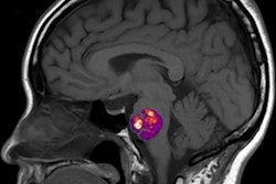
Trained using 414 patients clinically diagnosed with Alzheimer's disease and normal controls in the ADNI, the researchers' fully convolutional neural network (CNN) generates color-coded disease probability maps after analyzing T1-weighted brain MR images. Next, a multilayer perceptron analyzes these disease probability maps to produce a binary classification of Alzheimer's disease status.
This model, as well as other versions that incorporated additional types of nonimaging data, were assessed on the ADNI dataset, as well as on three independent cohorts of patients. These included 382 subjects in the Australian Imaging, Biomarker, and Lifestyle Flagship Study of Aging (AIBL), 102 participants in the Framingham Heart Study, and 582 subjects in the National Alzheimer's Coordinating Center (NACC).
The researchers found that the best-performing version of the model included consideration of nonimaging variables such as a patient's age, gender, and Mini-Mental State Examination (MMSE) score.
| Performance of AI analysis of MRI and nonimaging factors for classifying risk of Alzheimer's disease | ||||
| ADNI study | AIBL | Framingham Heart Study | NACC | |
| Area under the curve | 0.996 | 0.974 | 0.876 | 0.954 |
In a head-to-head comparison with a multi-institutional team of 11 practicing neurologists on a randomly selected set of 80 ADNI cases, the model produced an area under the curve (AUC) of 0.996, compared with an average AUC of 0.920 by the neurologists.
In other results, the model's identification of high-risk cerebral regions closely tracked postmortem histopathological findings, according to the researchers.
"If confirmed in clinical settings, this approach has the potential to expand the scope of neuroimaging techniques for disease detection and management," the authors wrote. "Further validation could lead to improved care and outcomes compared with current neurological assessment, as the search for disease-modifying therapies continues."
This type of approach has wide-reaching potential, especially in resource-limited settings, according to senior author Vijaya Kolachalama, PhD.
"Not only can we accurately predict the risk of Alzheimer's disease but this algorithm can generate interpretable and intuitive visualizations of individual Alzheimer's disease risk en route to accurate diagnosis," Kolachalama said in a statement.