A new type of elastography based on optical coherence tomography may one day help surgeons identify residual cancer in the margins during breast-conserving surgery, according to a study published in the April edition of Cancer Research.
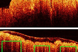
Quantitative microelastography (QME) builds on optical coherence tomography (OCT) technology, which creates images based on the amount of backscattered light, similar to how ultrasound creates images from sound waves. Researchers believe that QME could be a promising tool to help surgeons identify residual cancer during surgeries, reducing the number of women who need repeat surgery due to inadequate surgical margins.
"This study is the first to determine the accuracy of QME, or any variant of optical elastography, for assessment of tumor margins in specimens excised during [breast conserving surgery]," wrote the authors, led by Kelsey Kennedy, PhD, from the Harry Perkins Institute of Medical Research at the University of Western Australia (Cancer Research, April 2020, Vol. 80:8, pp. 1773-1783).
At first, OCT looked like a promising technology to help reduce residual cancer following breast-conserving surgery. But researchers found its high sensitivity and specificity for high-grade tumors didn't translate well to surgical margins.
The authors hypothesized that QME might overcome some of OCT's limitations. They put their theory to the test in a small feasibility study using fresh tissue samples from patients who had just undergone breast-conserving surgery.
Within 1 hour of excision, the researchers scanned the tissue samples using a benchtop wide-field QME system, which is in turn based on a spectrum-dominated OCT system. Seven readers, including surgeons and a medical sonographer, read the images.
Out of the 154 regions of interest in the study, 15.6% were confirmed to have cancer within 1 mm of the surface. Using QME, all seven readers correctly identified benign and malignant margins in the vast majority of cases.
The modality was associated with a 92.9% sensitivity, 96.4% specificity, and 95.8% accuracy. For comparison, OCT alone had a sensitivity, specificity, and accuracy of 69%, 79%, and 77.5%, respectively.
While QME looks like a promising technology for identifying cancer in surgical margins, the technology to implement it during surgery using a handheld device doesn't yet exist. Some members of the scientific community are working to develop handheld probes and image processing algorithms that may one day make intraoperative cancer detection a reality.
"The use of intrinsic tissue contact without need for exogenous dyes and the optimal trade-off in speed, field of view, and resolution provided by QME make it a promising candidate for improving intraoperative guidance of [breast conserving surgery]," the authors concluded. "More broadly, QME may be applicable in a range of surgical or preoperative biopsy guidance applications, particularly in cancers that are known to exhibit altered mechanical properties."