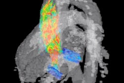
An investigational 4D flow MRI sequence has helped researchers confirm different blood flow characteristics in the hearts of young, healthy men and women. The findings could help clinicians set benchmarks for comparisons with unhealthy patients, according to a study published February 27 in Radiology: Cardiothoracic Imaging.
The MRI views of heart function revealed significantly higher peak systolic blood flow in the left ventricle of men, compared with women. Conversely, the females' hearts reached higher vorticity, which measures blood flow rotation during different points in the cardiac cycle. By establishing quantitative normal standards for both genders, the researchers hope to improve the assessment of cardiac performance in patients with suspected heart problems.
"Taken together, our results agree well with many of the previous findings of sex-dependent differences in cardiovascular function," wrote lead author David Rutkowski, PhD, and colleagues at the University of Wisconsin in Madison. "However, the major contribution of this work is that it is the first to use 4D flow MRI and cardiac strain analysis to determine sex differences in myocardial function in a young healthy adult population."
It is no secret that women's hearts are smaller in size and on average beat faster than men's hearts. However, information is limited on the differences between the genders in regard to blood flow, the amount of kinetic energy expended during contraction and filling of the left ventricle, and blood rotation in the ventricles.
Recently, 4D flow MRI has "gained traction in the evaluation of cardiac function in flow analysis," the authors noted. By analyzing blood velocity and rotation, the approach could help determine cardiac characteristics, such as kinetic energy and vorticity, and, "therefore, can be used with flow parameters to provide ventricular efficiency indexes."
To explore these factors in men and women, the researchers retrospectively analyzed data from 20 male (mean age, 25.8 ± 2.7) and 19 female (mean age, 27.1 ± 2.9) healthy volunteers. The 39 subjects underwent 3-tesla MRI scans (MR750 and Signa Premier, GE Healthcare) to measure left ventricle flow.
The 4D flow MRI parameters included 1.25-mm isotropic spatial resolution, 320-mm field-of-view, 6.2-msec repetition time, 160 cm/sec velocity encoding, and 11-minute scan time. Kinetic energy and vorticity metrics then were plotted across the group to determine average cardiac cycles and peak flow within the genders.
Rutkowski and colleagues found statistically significant differences in several comparisons. Men had greater peak systolic blood kinetic energy than women, while the females had greater peak diastolic vorticity indexes and cycle-average vorticity in the left ventricle than men.
| Gender comparison of blood flow performance | |||
| Men | Women | p-value* | |
| Peak systolic blood KE | 4.76 mJ | 3.36 mJ | 0.047 |
| Peak systolic vorticity index | 0.008 | 0.014 | 0.007 |
| Cycle-average vorticity | 0.006 | 0.011 | 0.001 |
*All p-values are statistically significant.
"Additionally, the female heart showed increases in myocardial global strain and strain rates (systolic and diastolic), which may be related to the increased left ventricle vorticity observed with 4D flow MRI in women," the authors wrote.
Rutkowski and colleagues currently are using 4D flow MRI to look at patients with atrial fibrillation, with the hope that the modality will detect patterns and relationships in the atria, the upper chambers of the heart, similar to those found in the ventricles.