Targeting joint capsule abnormalities in the shoulder on MRI scans can help determine which patients with rotator cuff tears will have preoperative shoulder stiffness, according to a study published February 19 in the American Journal of Roentgenology.
With the ability to predict which patients will have a limited range of motion from the rotator cuff injury, physicians can better decide on the most effective course of therapy or surgery.
"The results show that MRI findings -- especially joint capsule edema and thickness at the axillary recess -- can be useful in predicting stiff shoulder in patients with rotator cuff tear," wrote the authors, led by Dr. Yoon Yi Kim, from the Veterans Health Service Medical Center in South Korea.
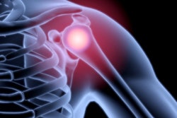
Estimates are that more than 40% of patients with a rotator cuff tear injury commonly deal with shoulder stiffness, which is clinically known as adhesive capsulitis and involves thickening and contraction of the shoulder joint capsule. Several factors, ranging from a full-thickness tear to a small labral tear and ligament damage, can contribute to shoulder stiffness. Patients with preoperative adhesive capsulitis also are likely to take longer to recover an adequate range of motion.
The challenge for physicians is that a physical exam cannot always determine the primary cause of adhesive capsulitis. Hence, MRI becomes the modality of choice to observe joint capsule abnormalities, ligament thickening, tears, and other shoulder anomalies. However, previous research has yet to determine an association between joint capsule abnormality on MRI and shoulder stiffness in patients with rotator cuff tears, Kim and colleagues noted.
"The current study was initiated by observing several cases showing rotator cuff tear and joint capsule abnormality on MRI in patients with clinical impression of adhesive capsulitis," the authors wrote. "We hypothesized that a joint capsule abnormality in patients with full-thickness rotator cuff tears could be a factor affecting shoulder stiffness."
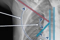
The researchers analyzed 106 patients (mean age, 71.2 ± 4.5 years; range, 59-85 years) with full-thickness rotator cuff tears of 5 cm or less. Shoulder MRI scans were performed on either a 1.5-tesla system (Signa HDxt, GE Healthcare) or 3-tesla system (Magnetom Skyra, Siemens Healthineers) with a dedicated shoulder coil. They looked for several shoulder abnormalities, including swelling and thickness in the joint capsule and degeneration of the torn rotator cuff muscle and correlated the MR images with patient's degree of shoulder stiffness, such as their inability to elevate their arms and their restricted range of motion.
Kim and colleagues indeed found significant correlations between patients' limited range of motion, particularly with forward elevation, and thickness of their joint capsule in the glenoid portion and axillary recess of the shoulder. In addition, swelling of the joint capsule in the humeral area of the shoulder was significantly associated with shoulder stiffness and lack of arm elevation.
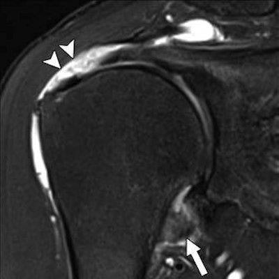
 MR images are from a 67-year-old woman with shoulder pain and limited range of motion on external rotation, forward elevation, and internal rotation. Patient underwent rotator cuff repair with arthroscopic capsular release. Above: Oblique coronal fat-suppressed T2-weighted MR image shows thickening and edema in glenoid and humeral portions of joint capsule at axillary recess (arrow). Note full-thickness tear of supraspinatus tendon (arrowheads). Below: Oblique sagittal proton density MR image shows partial obliteration of subcoracoid fat triangle (arrow). Images courtesy of the AJR.
Given the finding that MRI can link joint capsule abnormality to shoulder stiffness in patients with rotator cuff tears, the researchers believe this information can be especially useful as a reference standard on how best to determine the most appropriate treatment for the condition.