Canadian researchers found that their artificial intelligence (AI) algorithm was able to find and quantitate infarcted brain tissue on baseline CT studies in patients with acute ischemic stroke, potentially helping to improve treatment decisions, according to research published online January 28 in Radiology.
Based on a combination of deep-learning and machine-learning techniques, an algorithm developed by a research team from the Calgary Stroke Program produces ischemic lesion volume calculations that correlate well with volumes calculated by experts on diffusion-weighted MRI (DWI-MRI).
"This work might be translated into clinical settings to help physicians make clinical decisions for these patients," wrote the authors, led by Wu Qiu, PhD, and Hulin Kuang, PhD.
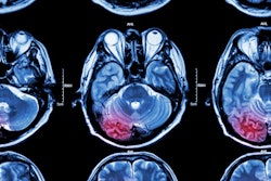
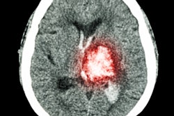
In patients with acute ischemic stroke, it's important to find and determine the extent of infarcted brain tissue on baseline scans performed when they are admitted; patients that have extensive infarction are unlikely to benefit from thrombolysis or thrombectomy procedures. The Alberta Stroke Program Early CT Score (ASPECTS) can be used as a semiquantitative approach to subjectively assess the extent of infarction on unenhanced CT images, but this method requires expertise and offers only an approximation of the total extent of early ischemic changes, according to the researchers.
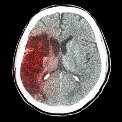
Quantitative estimates of infarctions on unenhanced CT are also challenging to perform. As a result, the researchers sought to develop an automated method by combining manually defined features with deep learning-based features in order to detect and quantify infarction on baseline unenhanced CT scans.
They first gathered data from 257 acute ischemic stroke patients from the Korean Stroke Registry who presented within eight hours of being well and who had undergone baseline unenhanced CT scans with a slice thickness of 5 mm or smaller, followed by DWI-MRI within one hour.
Of these 257 patients, 157 were randomly selected for algorithm training and 100 were set aside for testing. After a fellowship-trained physician manually segmented the ischemic lesions on the DWI-MRI scans, these expert-contoured early ischemic change lesion labels were used for training and testing of the model. The DWI-MRI lesion volume was determined by multiplying the number of voxels within the manually contoured lesion region by the resolution of the DWI-MRI scan, according to the researchers.
A convolutional neural network was then utilized to provide a probability map associated with degree of hypoattenuation for each voxel. This map and four other features were used to train a random forest classifier machine-learning model.
In testing, the algorithm-detected lesion volume correlated well with the reference standard of expert-contoured lesion volume on acute DW-MRI exams (r = 0.76, p < 0.001). In addition, the mean difference between the two methods was 11 mL, a difference that was not statistically significant (p = 0.89).
"If its potential is realized in a broader setting, this machine-learning method will provide a key imaging element required for treatment decision-making," noted Dr. Kambiz Nael of the Icahn School of Medicine at Mount Sinai in an accompanying editorial. "In that case, the simplicity of noncontrast-enhanced CT would rival information obtained with either CT perfusion or MRI."