CHICAGO - MRI brain scans can play a valuable role in the discovery and evaluation of abnormalities in the brains of people with depression, potentially leading to effective treatments for the disorder, according to two studies presented this week at RSNA 2019.
In both presentations, researchers tested their newly developed MR imaging protocols to drill deeper into the reasons for major depressive disorder. With a better understanding of the neurological reasons for the condition, better treatment might help alleviate the symptoms and resultant distress.
"Unfortunately, with current treatments, there is a large chance of relapse or recurrence," said Kenneth Wengler, PhD, from Columbia University in New York City, in a statement. "To develop new, more effective treatments, we must improve our understanding of the disorder."
In the first study, co-author Wengler and colleagues from Stony Brook University in Stony Brook, NY, evaluated their newly developed MR imaging technique called intrinsic diffusivity encoding of arterial labeled spins (IDEALS), which targets the blood-brain barrier and the movement of water out of the blood vessels and into the brain tissue.
A total of 14 patients with major depressive disorder and 14 healthy control subjects underwent MRI brain scans using the IDEALS protocol. The images showed less water moving from inside the blood vessels to the outside in patients with the disorder, which would indicate a disruption in blood-brain barrier integrity. This difference was particularly significant in the gray matter regions of the amygdala and the hippocampus, which are associated with emotions and memory, respectively.
"We observed disruption of the blood-brain barrier in gray matter regions known to be altered in major depressive disorder," Wengler said. "This study helps improve our understanding of the pathophysiology of depression and can open new avenues of treatment for a disorder that affects over 100 million individuals worldwide."
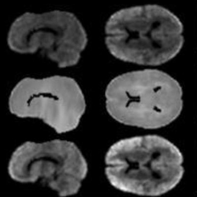
 MRI brain scans using the newly developed IDEALS technique shows the difference between normal brains and patients with major depressive disorder. Images courtesy of RSNA.
In the second study, researchers at the University of North Carolina (UNC) used resting-state functional MRI (fMRI) to evaluate 66 adults with major depressive disorder and 66 matched healthy controls with a newly developed multiscale neural model inversion approach designed to link the brain's microscopic circuitry with larger-scale interactions.
"Current methods of studying the brain provide a superficial understanding of connectivity," said study co-author Guoshi Li, PhD, a research associate at UNC, in a statement. "This method allows us to identify impaired connectivity within each brain region, making it a potentially more powerful tool to study the neuromechanism of brain disorders and develop more effective diagnosis and treatment."
As part of the study, Li and colleagues analyzed the excitatory or inhibitory influence between neuronal cell groups. A proper balance between these two brain functions is crucial to a person's well-being. In fact, previous research has indicated that poor inhibitory control over the amygdala could cause depressive symptoms.
In this case, the fMRI results identified abnormal patterns of excitation and inhibition at the dorsal lateral prefrontal cortex in patients with major depressive disorder. Both brain regions are associated with cognitive control and proper function of the amygdala.
"In our study, we found that excitation and inhibition in the brain regions in control of executive functions and emotional regulation were reduced in patients with major depressive disorder," Li added. "This suggests that control functions in major depressive disorder are impaired, which may lead to elevated responses in the amygdala, resulting in increased anxiety and other negative moods."
The UNC researchers also found that recurrent excitation in the thalamus region of the brain, which also is involved in the control of emotions, was abnormally high in patients with major depressive disorder.
The findings from both studies could open the door for a deeper understanding of the mechanisms behind depression and potentially better ways to treat the condition.