Contrast-enhanced digital mammography (CEDM) and molecular breast imaging (MBI) are comparable to breast MRI when it comes to identifying breast cancer, but CEDM and MBI outperform breast MRI when it comes to treatment staging, according to a study published online October 29 in Radiology.
The findings demonstrate that CEDM and MBI are valuable tools in the diagnostic arsenal, offering clinicians an alternative to breast MRI, wrote a team led by Dr. Jules Sumkin of the University of Pittsburgh Medical Center.
"Our results showed that CEDM and MBI are effective in the local staging of disease in women with newly diagnosed breast cancer, with similar visualization of index cancers and higher specificity for additional malignancy when compared with MRI," the researchers wrote.
Dynamic contrast-enhanced (DCE) MRI is often used to stage newly diagnosed breast cancer, but it does have its drawbacks, including limited access, high cost, and false-positive findings, Sumkin and colleagues noted. That's why the team investigated the efficacy of CEDM and MBI, comparing the performance of the three modalities for identifying breast cancer -- using pathology as the standard -- as well as their positive predictive value for additional biopsies.
The study included data from 99 women with 110 biopsy-proven breast cancers who underwent breast MRI, CEDM, and MBI between October 2014 and April 2018. Eight radiologists read the exams, blinded to interpretations of each modality. The readers compared the visibility of index cancers, lesion sizes, and additional suspicious lesions among the three exams.
All three modalities demonstrated comparable performance when it came to identifying cancer: Breast MRI found 102 of the 110 (93%), CEDM found 100 (91%), and MBI found 101 (92%).
But CEDM and MBI outperformed breast MRI in the following ways:
- Breast MRI overestimated the size of malignant lesions by 24%, while CEDM overestimated by 11% and MBI overestimated by 15%.
- Breast MRI found almost twice as many suspicious lesions (46) compared with CEDM (27) and MBI (25), but it did not identify additional cancers.
- Breast MRI resulted in a lower positive predictive value of additional biopsies for breast MRI (28%) compared with CEDM (52%) and MBI (44%).
"Both CEDM and MBI showed visualization of index malignancies similar to that of MRI but less frequently led to overestimation of lesion size ... [and] both CEDM and MBI produced fewer additional suspicious findings than did MRI while depicting similar numbers of nonindex malignancies," Sumkin and colleagues wrote.
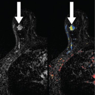
 Left: A 48-year-old woman with a 1.3-cm invasive ductal carcinoma (arrow) in the right breast at MRI with color mapping. Right: A 0.5-cm mass (arrow) was seen in the left breast that was biopsy-proven benign. Neither was seen at MBI or CEDM. Images courtesy of Radiology.
Having CEDM and MBI in one's clinical toolbox offers an alternative for assessing the extent of disease in women who cannot tolerate breast MRI, the group concluded.
"Because it is still early in terms of our experience, MRI remains our primary modality in the local preoperative staging of breast cancer," the team wrote. "However, as a direct result of this study, we have begun incorporating CEDM as an alternative to MRI in evaluating the extent of disease in patients who cannot undergo MRI."