How much easier would it be to image a knee with a stretchable, wearable MRI knee coil? Swiss researchers are working on this technology for musculoskeletal (MSK) MR imaging, and they reported early success in their work at the recent International Society for Magnetic Resonance in Medicine (ISMRM) annual meeting in Montreal.
Current conventional MRI coils are rigid and do not mold well to patient anatomy. This deficiency compromises sensitivity in MSK MR imaging for some patients and reduces comfort for others. But it doesn't have to be that way.
"Imagine a coil that would ... adapt to the patient," said device co-developer Andreas Port, PhD, from the Institute for Biomedical Engineering at ETH Zürich. "We believe such a coil would be stretchable and elastic and would conform very well to the body part being imaged. This would offer good sensitivity and good patient comfort at the same time."
Relaxed fit
Researchers around the world have been working on adjustable, flexible MRI coil designs for many years, but finished products typically maintain a degree of rigidity due to components such as copper wiring. The results are not always a perfect fit for all patients.
"We all come in different shapes and sizes, as do the patients who come to our MR centers," Port told ISMRM 2019 attendees. "And yet, what they find there is MR scanner equipment that is a fixed size."
Thus, the need exists for more adaptable devices to allow for a wider range of clinical approaches to image MSK joints and muscles at various angles and/or while they are in motion. Technical challenges arise, however, in making MRI coils more adaptable to a patient's configuration.
While copper offers high electrical conductivity to produce good imaging performance, it is not flexible or stretchable at all, which is what would be required for a better-contoured coil. Previous work in this area has explored the potential of flexible coils through multiple-resonator conductors and high-impedance coils. Among the forward-thinking developers are researchers from NYU Langone Health who are working on an MRI device that fits over a patient's hand like a glove to image moving joints.
Liquid metal
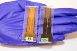
For this Swiss project, Port and colleagues began with gallium indium, a metal alloy that transforms to liquid at room temperature (60° F), offers high electrical conductivity, and has low toxicity. The gallium indium was injected into a stretchable silicone tube, which then was placed into a copper insert and heat-shrink tubing to seal the liquid metal in place and to provide electric conductivity. The result was a highly stretchable MRI coil.
 After liquid gallium indium is injected into a silicone tube, electrical contact is formed by inserting a copper wire (a) into the tube and sealed by instant adhesive and a heat-shrink tube. Using the same manufacturing process, coils with tubes of different inner and outer diameters (b) can be produced. The coil is highly stretchable, lightweight, and easy to handle. Images courtesy of ISMRM.
For optimum MR imaging, coil conductivity must have the least resistance possible. Gallium indium has high electrical conductivity; a coil made of this liquid metal will have a resistance greater than that of copper at 0 MHz. At a higher frequency of 128 MHz, a phenomenon known as the skin effect occurs, which improves the flow of electrical current in and out of the external layer of the silicone tube, Port explained. The enhanced flow reduces gallium indium resistance from 17 times that of copper to four times, making the MRI coil more efficient.
To adapt this technology for clinical application, the researchers combined several stretchable elements into a wearable coil array. In the first iteration, the coil array was attached to stretchable textile material. They then added a stretchable material to create an outer layer, or casing, into which the coil element was inserted.
"This is a very specific application and could offer very interesting functional insights," Port added. "A wearable coil also could be put on by the patient him or herself just like a piece of clothing outside of the scanner room, improving workflow."
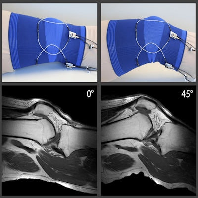
To test the technology, the researchers tried their stretchable MRI coil on a 3-tesla MRI scanner (Ingenia, Philips Healthcare). At various angles, the resultant MR images showed the interaction of the knee, thigh bone, and cruciate ligaments and how they interact as the knee flexes. The researchers also performed a dynamic knee study, during which a volunteer moved his knee freely inside of the scanner. Again, the exercise was successful in showing how knee movements affect the cruciate ligaments.
 In vivo MR images are from a healthy volunteer's knee at two different flexion angles. The array was tuned and matched for the 0° position only, and no further adjustments were made for the 45° position. Images courtesy of ISMRM.
In evaluating the early results, the researchers wrote in their ISMRM paper that the stretchable tube coils achieved "adequate mechanical and electrical robustness as well as sealing performance" and "do not appear to suffer from chemical deterioration, such as oxidation and corrosion." The array coil also is "extremely flexible and lightweight," which would be advantageous for patient evaluation and comfort.
"In the future, such a wearable coil could be complemented with variable detection, eliminating cabling to the array by on-body digitization and MR signal transmission by in-optical link," Port added. "Altogether, I believe a wearable coil technology may take us a step forward toward MR systems that adapt to the patients and not the other way around."