Chemotherapy performed on women during pregnancy could adversely affect long-term cortical development in their children's brains, according to a preliminary study presented last week at the International Society for Magnetic Resonance in Medicine (ISMRM) annual meeting in Montreal.
Belgian researchers found significantly decreased cortical thickness in the left precentral sulcus and increased gyrification in the left postcentral sulcus among children at age 9 whose mothers underwent chemotherapy after their first trimester, compared with healthy control subjects. These particular areas of the brain are associated with attention and executive function.
"These are very subtle effects, and on their own are not a contraindication not to treat these women during pregnancy," lead author Jeroen Blommaert, PhD, from the department of oncology at KU Leuven, said of the preliminary results. "We need to be more sure of the effects of this therapy during pregnancy, and we need to gather more data through a longer follow-up period."
Estimates are that one in every 1,000 to 2,000 pregnant women will be diagnosed with cancer, and increasingly, chemotherapy is recommended for treatment during the pregnancy. Today, approximately 71% of women with maternal cancer are treated with timely chemotherapy during pregnancy, Blommaert told ISMRM attendees.
Chemotherapy is contraindicated during the first trimester due to an increased risk of congenital anomalies, but the treatment can be administered during the second and third trimesters. Previous studies have shown that this exposure in utero does not add to the risk of congenital malformations, but evidence suggests that it could adversely affect a child's cognition, and a greater number of children are born late or premature.
"We also see a higher amount of these children being born small for their gestational age," Blommaert added. "This leads to the current practice to try to elongate the pregnancy as long as possible in favor of [cancer] treatment during this time."
Data are very limited, however, because past research has followed these children for only up to three years, and few researchers have studied the potential long-term effects of chemotherapy on the neurological development of children.
To study the issue further, researchers analyzed 16 children at 9 years of age whose mothers underwent chemotherapy after the first trimester of their pregnancies; they compared them to 29 healthy controls who were matched by age, gender, and prematurity at birth. All of the subjects underwent 3-tesla MRI scans (Achieva, Philips Healthcare) with a 32-channel phased-array head coil.
For this ISMRM presentation, Blommaert focused on results from T1-weighted axial 3D magnetization-prepared rapid acquisition gradient-echo (MP-RAGE) images to show differences in cortical thickness. The researchers also calculated a gyrification index to show how formation of the cerebral cortex may have been altered.
The MR images revealed significantly reduced cortical thickness in the left precentral sulcus among children whose mothers underwent chemotherapy, compared with the healthy control group. Conversely, the team noted a significant increase in the gyrification index in the left postcentral sulcus among the children exposed to chemotherapy, which could indicate a cortical malformation or subclinical tendency for polymicrogyria, which is associated with seizures.
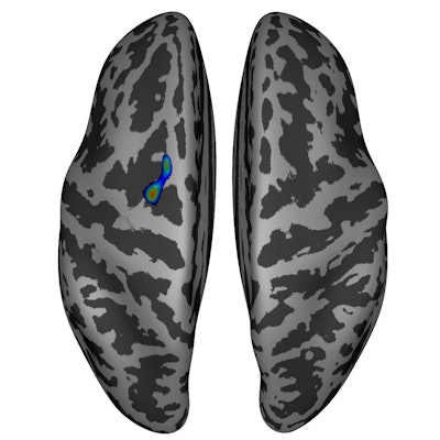
 Significantly reduced cortical thickness (p < 0.05) was found in the superior part of the left precentral sulcus (shades of blue, green, and red) in children prenatally exposed to chemotherapy. Results are corrected for gestational age. Image courtesy of the ISMRM.
"To our knowledge, this is the first study to investigate brain development after prenatal exposure to chemotherapy," Blommaert and colleagues wrote in their ISMRM abstract. "The findings suggest that prenatal exposure to chemotherapy impacts cortical development through both thickness and gyrification, possibly influencing attentional development. These results need validation in a larger sample, which is currently being collected."