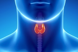
Using a combination of advanced ultrasound techniques improves the accuracy of the modality for diagnosing suspicious thyroid nodules compared with conventional ultrasound alone, according to a study published online March 1 in the International Journal of Clinical Oncology.
Chinese researchers used a combination of superb microvascular imaging (SMI), real-time elastography (RTE), and conventional ultrasound to analyze the characteristics of thyroid nodules that could indicate malignancy, wrote a team led by Dr. Shufang Pei of Southern Medical University, Guangzhou, Guangdong.
"The ultrasonographic features of these thyroid nodules are complex, and the characteristics of conventional ultrasound images often overlap," the group wrote. "The conventional ultrasound features of some nodules were shown to be benign, and the pathology of the surgery was confirmed to be malignant; while the conventional ultrasound features of some nodules showed malignancy, and the pathology confirmed by surgery was benign. ... Therefore, it is particularly important to accurately diagnose benign and malignant nodules."
Pei and colleagues conducted a study that included 196 thyroid nodules classified as category 4 using the Thyroid Imaging Reporting and Data System (TI-RADS 4) -- first imaged on ultrasound alone, then with a combination of ultrasound, SMI, and RTE. The group compared the sensitivity, specificity, accuracy, false-negative rate, and false-positive rate of each ultrasound method; the gold standard was surgical pathology results.
Of the nodules imaged, 78 were benign and 118 were malignant. The researchers found that their combination technique improved the diagnostic accuracy of TI-RADS 4 nodules and also helped differentiate malignant from benign nodules.
| Advanced ultrasound techniques vs. conventional ultrasound for thyroid nodules | ||||
| Performance measure | Ultrasound alone | Superb microvascular imaging (SMI) | Real-time elastography (RTE) | Ultrasound + SMI and RTE |
| Sensitivity | 65.3% | 78% | 80.5% | 94% |
| Specificity | 69.2% | 93.6% | 84.6% | 87.2% |
| Accuracy | 66.8% | 84.2% | 82.1% | 91.3% |
| False-negative rate | 34.8% | 22% | 19.5% | 6.9% |
| False-positive rate | 30.8% | 6.4% | 15.4% | 12.8% |
The sensitivity, specificity, and accuracy rates of SMI and RTE were higher than those for ultrasound alone. Additionally, the group found that RTE had a higher sensitivity than SMI but also a higher false-positive rate.
It was the combination of the three that was the winning ticket.
"The results of this study suggest that the application of RTE and SMI may help compensate in areas in which conventional ultrasound may be deficient in assessing the TI-RADS category 4 nodules," the group concluded. "Thus, multimodal ultrasound imaging -- using the three methods -- may provide more comprehensive information regarding the nodules, facilitating more accurate diagnoses."