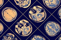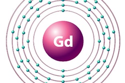
Researchers have discovered the presence of gadolinium from MRI contrast in gliomas, as well as neighboring normal brain tissue and necrosis. The findings are contributing to the body of knowledge regarding the retention of trace elements from gadolinium-based contrast agents (GBCAs), according to a study published online February 2 in Neuroradiology.
Researchers from Finland found that gadolinium deposits were highest in gliomas with noticeable contrast enhancement and in patients who received a linear GBCA, rather than a macrocyclic GBCA. Similar retention levels and the influence of GBCA type also were evident in normal brain tissue and necrosis samples.
"To our knowledge, this is the first study to provide quantitative data of gadolinium retention in gliomas and neighboring normal brain with respect to tumor enhancement and type of GBCA used," wrote the researchers, led by Dr. Aida Kiviniemi from Turku University Hospital. "The levels of gadolinium in the tumor and normal brain suggest a possible transit of gadolinium to the surroundings of the brain lesion."
Quantitative data
Much has been researched and written about the retention of gadolinium in recent years and its association with linear and macrocyclic GBCAs long after contrast administration. Unlike previous studies, the current research was designed to provide quantitative data of gadolinium retention relative to gliomas and adjacent normal brain tissue, as well as information on how the type of GBCA administered might play a role.
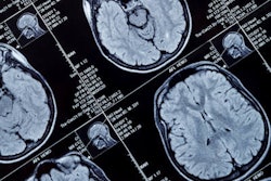
The retrospective study included 69 patients with newly diagnosed primary glioma who underwent contrast-enhanced MRI before surgery. Seven patients received a linear GBCA -- either gadodiamide (Omniscan, GE Healthcare) or gadopentetate dimeglumine (Magnevist, Bayer HealthCare) -- while the other 62 patients received a macrocyclic GBCA -- either gadobutrol (Gadovist, Bayer) or gadoterate meglumine (Dotarem, Guerbet).
Gadolinium deposition was measured from histologically viable tumor samples in all 69 patients, along with 13 normal brain tissue samples and necrosis samples from 14 glioblastomas. The amount of tumor GBCA enhancement was assessed as none, minimal, or noticeable. The researchers also noted differences in gadolinium retention relative to the type of GBCA.
Gadolinium was detected in 39 (57%) glioma tumor samples, eight (62%) normal brain samples, and 12 necrotic specimens. The linear GBCAs gadodiamide and gadopentetate dimeglumine resulted in significantly higher tumor gadolinium presence, compared with the macrocyclic GBCA gadoterate meglumine (p < 0.01 and p < 0.05, respectively).
Of the eight patients with gadolinium in normal brain tissue, two received the linear GBCA gadodiamide, while six were administered the macrocyclic GBCA gadoterate meglumine. Gadolinium concentration in the normal brain tissue was significantly higher (p = 0.05) after exposure to linear gadodiamide, compared with macrocyclic gadoterate meglumine. Gadolinium exposure within necrosis samples was similarly significantly higher (p = 0.05) with linear gadodiamide, compared with macrocyclic gadoterate.
While contrast enhancement in brain tumors is expected, how and why is gadolinium found in adjacent normal brain tissue where contrast uptake is less likely? Previous studies have suggested that cerebrospinal fluid or the brain's glymphatic system might provide the pathway.
"The exact mechanism behind the retention ... remains unsolved," Kiviniemi and colleagues wrote. "A small but significant correlation between the amount of gadolinium in the tumor and normal brain was detected, implying passage of gadolinium from the site of pathologic brain lesion. While further studies are needed to confirm this association, an interesting question arises whether gadolinium traverses from the region of brain lesion to neighboring sites in other brain abnormalities such as ischemia, infection, or multiple sclerosis."