Proton MR spectroscopy (MRS) scans of newborns suspected of having brain damage can hasten an early and accurate diagnosis and help determine if they will have severe developmental issues within two years after they are born, according to a study published online on November 14 in Lancet Neurology.
The key is MR spectroscopy's ability to measure the level of the compound N-acetylaspartate (NAA) in the baby's thalamus, which can be severely damaged by a loss of oxygen. Oxygen deprivation causes neonatal encephalopathy, which hampers brain development and neurological functions.
High levels of NAA in the thalamus indicate normal brain function, while low levels indicate brain damage. In the current study, proton MR spectroscopy predicted adverse outcomes at two years with 98% accuracy, based on initial NAA results.
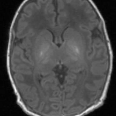
 MR spectroscopy shows the brain of a baby with brain damage. There is abnormal brightness in the deep nuclei in the center of the brain. Image courtesy of Imperial College London.
"Proton MR spectroscopy should be a routine component of clinical MR protocols for infants with neonatal encephalopathy," wrote lead author Dr. Peter Lally from Imperial College London and colleagues. "This assessment is ideal for early prognostication and might provide promising surrogate outcome measures for multicenter clinical trials. If early-phase trials were to employ proton MRS measures, such as NAA concentration, this would greatly increase their power to detect important treatment effects while potentially reducing their duration by several years."
When newborns are suspected of having brain damage, the first course of action is typically an MRI scan. However, previous research has found that MRI's accuracy for diagnosis ranges from 60% to 85%. With such mediocre results, it may take until the child reaches 2 years of age before physicians can confirm brain damage from birth.
In comparison, thalamic proton MRS performed within a few days or weeks of birth has been shown to provide insight into the metabolic effects of neonatal encephalopathy.
To assess the value of MRS in this patient population, the researchers conducted a prospective, multicenter study between January 2013 and June 2016 in eight neonatal intensive care units in the U.K. and U.S. They included 223 term and near-term neonates who received treatment for neonatal encephalopathy. The babies underwent T1-weighted imaging, T2-weighted imaging, diffusion-weighted MRI, and thalamic proton MR spectroscopy four to 14 days after birth. MRI scans were performed on 3-tesla scanners (Philips Healthcare, Siemens Healthineers, or GE Healthcare).
In addition, 190 (85%) of the infants received clinical neurodevelopmental tests 18 to 24 months after birth. The primary outcome was the association between the MR results and death or moderate or severe disability.
At two years, 31 (16%) of the followed-up children had moderate or severe disability, including one death. MRS-derived thalamic NAA results were proficient in predicting an adverse neurodevelopmental outcome, with sensitivity of 100%, specificity of 97%, and accuracy of 98%.
"The results provide evidence of the high accuracy of thalamic proton MR spectroscopy, acquired days after birth, in predicting adverse neurodevelopment two years after neonatal encephalopathy," the researchers concluded. "Thalamic NAA concentration had the highest accuracy in predicting neurodevelopmental outcome following neonatal encephalopathy and was associated with performance across cognitive, language, and motor domains at 2 years of age."