With the help of MRI, ophthalmologists in the U.K. were able to discover what was causing eyelid swelling in a 42-year-old woman: a hard contact lens that had been embedded in her eye for 28 years. The bizarre case was published on August 10 in BMJ Case Reports.
The woman was referred to ophthalmology from her general practitioner after complaining of swelling in one of her eyes. The swelling began as a nontender, pea-sized lump that grew larger and became painful to the touch, reported a team led by Dr. Sirjhun Patel of the National Health Service Tayside in Dundee, Scotland. The woman also reported experiencing ptosis, or drooping of the upper eyelid, which she said had never bothered her.
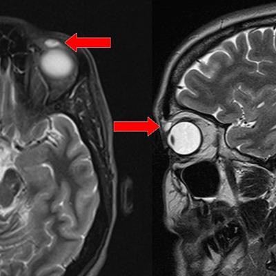
 T2-weighted MRI scan of the head: transverse and sagittal views. Red arrows show high-intensity signal nodular lesion in the left upper eyelid. Image courtesy of BMJ Case Reports.
An MRI scan of the orbit was performed, and it revealed a well-defined ovoid nodule measuring 8 x 4 x 6 mm. Features on the MRI scan were consistent with a cyst with proteinaceous content, Patel and colleagues reported. There were no other features of any foreign body.
The nodule was excised and found to be an encapsulated cyst. Upon further investigation, the cyst ruptured and the clinicians extracted a rigid gas permeable (RGP) contact lens.
The woman was questioned further, and she told clinicians that at the age of 14 she had been hit in the upper left eyelid with a shuttlecock while playing badminton. She was wearing a hard contact lens in the eye at the time, but she never found the lens and assumed it was dislodged from the eye and lost, according to the authors.
"We can infer that the RGP lens migrated into the patient's left upper eyelid at the time of the trauma and had been in situ for the last 28 years," they wrote.
Patel and colleagues discovered one other case of a contact lens that had migrated and had a similar long-term period of retention. But it was still a mystery in the current case as to why the area around the lens began swelling nearly three decades later.
They credited MRI with a radiofrequency surface coil as key to helping them detect the lens, thanks to the imaging modality's submillimeter resolution.
"This alternative method of imaging may have provided better visualization of the eyelid anatomy and possibly identified the foreign RGP lens in situ," they concluded.