Functional MRI (fMRI) scans have helped open researchers' eyes to which brain regions provide the primary connections and associations between poor sleep quality and depression, according to a study published online July 25 in JAMA Psychiatry.
The findings provide a "neural basis for understanding how depression is associated with poor sleep quality, and this in turn has implications for treatment because of the brain areas identified," wrote lead author Wei Cheng, PhD, from Fudan University in Shanghai, China, and colleagues in the U.K.
While it has been known for some time that poor sleep quality can lead to depression, knowledge about the neural connections that affect both conditions is lacking. Being able to target the culprit brain regions could advance treatments for depression and related sleep problems.
For this study, Cheng and colleagues analyzed data from 1,017 participants (546 female [54%]; age range, 22-35 years) drawn from a general U.S. population and who were enrolled in the Human Connectome Project (HCP).
Of the subjects, 92 people (9%) had been diagnosed with a major depressive episode during their lives and had significantly higher mean scores (7.55 ± 4.83 points) on the Depressive Problems scale than the other participants (3.70 ± 3.07) (p < 0.001).
As part of the project, all participants received resting-state fMRI scans on a 3-tesla system (Magnetom Skyra, Siemens Healthineers) and self-reported their depression issues and sleep quality patterns. The fMRI data were then used to segment gray matter of the whole brain into 250 regions of interest for more detailed evaluation.
In addition, to further validate the authenticity of the sleep and depression data, the researchers compared the HCP findings with results from 8,718 participants in the U.K. Biobank.
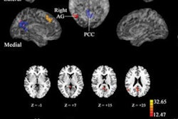
Cheng and colleagues discovered 162 functional connections involving areas associated with sleep. The key regions included the precuneus, anterior cingulate cortex, and lateral orbitofrontal cortex. Of those connections, 39 areas also were associated with the Depressive Problems scores.
Most important, the researchers found 11 brain areas with increased functional connectivity associated with both sleep and depression: the lateral orbitofrontal cortex, dorsolateral prefrontal cortex, anterior and posterior cingulate cortices, insula, parahippocampal gyrus, hippocampus, amygdala, temporal cortex, and precuneus.
As a result, Depressive Problems scores significantly correlated with poor sleep quality (r = 0.371, p < 0.001). In addition, a mediation analysis further confirmed the significant role those key brain regions play in the association between depressive problems with poor sleep quality (p < 0.001).
"A strength of this investigation is that the effects are likely to be very robust, given the large sample of participants with resting-state fMRI and the cross-validation with the U.K. Biobank dataset with a sample of 8,718 participants in one of its first uses of resting-state fMRI data," Cheng and colleagues noted.