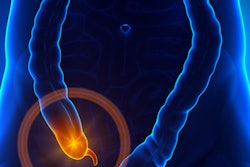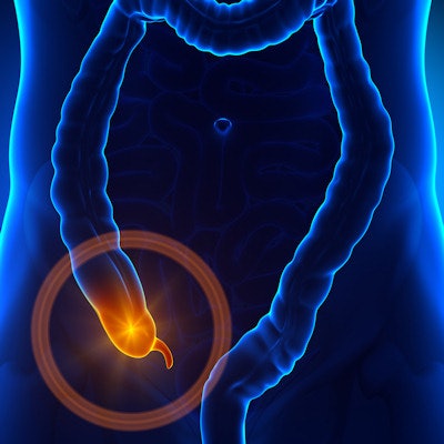
When used to diagnose and confirm cases of acute appendicitis in the emergency department (ED), MRI can provide the same efficacy as CT without the risk of radiation exposure for patients, according to a study published in the August issue of Radiology. The findings support the use of MRI first for some patients.
The study was restricted to patients who were not pregnant and who were at least 12 years old. In this group, the researchers generally found no significant differences between CT and MRI in terms of diagnostic accuracy for acute appendicitis.
"Based on these results, we propose that intravenous contrast-enhanced MR imaging may be a viable initial imaging option for the evaluation of appendicitis in the ED in nonpregnant patients," wrote lead author Dr. Michael Repplinger, PhD, and colleagues from the University of Wisconsin-Madison.
Concern over exposure to radiation from CT scans has increased in recent years as clinicians balance the risks and benefits for patients. MRI is seen as a viable option, especially given recent studies indicating that diagnostic accuracy is equivalent between MRI and CT in appendicitis cases.
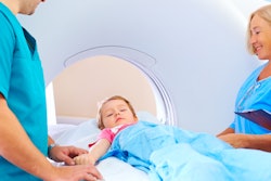
The researchers therefore decided to perform a prospective study that included 198 patients (mean age, 31.6 ± 14.2 years) who were evaluated for appendicitis between February 2012 and August 2014. They specifically recruited patients at least 12 years of age to minimize the need for sedation. There were 64 cases (32%) of appendicitis within the group.
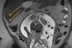
All patients received contrast-enhanced CT scans of the abdomen and pelvis on a 64-slice scanner (GE Healthcare). After CT, the patients underwent the research MRI protocol on a 1.5-tesla system (Signa HDxt or Discovery MR450w, GE) with an eight-channel or 12-channel phased-array body coil (Radiology, August 2018, Vol. 288:2, pp. 467-475).
Three radiologists interpreted the images and set a confidence level of 3 or greater as an indication of a positive result for appendicitis. The primary outcome was based on sensitivity and specificity of MRI for diagnosing the patient's condition.
In comparing the modalities' performances, the only statistically significant differences were observed in CT outpacing MRI in specificity and positive predictive value (PPV) when the trio of radiologists rated the likelihood of appendicitis at 3 or greater. When the likelihood was 4 or greater, there were no statistically significant differences between the modalities.
| CT vs. MRI for acute appendicitis, by confidence level | |||
| CT | MRI | p-value | |
| Confidence of 3 or higher | |||
| Sensitivity | 98.4% | 96.9% | 0.564** |
| Specificity | 89.6% | 81.3% | 0.012* |
| Positive predictive value | 81.8% | 71.3% | 0.010* |
| Negative predictive value | 99.2% | 98.2% | 0.511** |
| Confidence of 4 or higher | |||
| Sensitivity | 98.4% | 96.9% | 0.564** |
| Specificity | 93.3% | 89.6% | 0.166** |
| Positive predictive value | 87.5% | 81.6% | 0.152** |
| Negative predictive value | 99.2% | 98.4% | 0.541** |
**Not statistically significant.
The researchers cited several limitations of the study, including its small sample size, which skewed subjects' mean age to a younger number and a higher prevalence of appendicitis. The study was also conducted at a single facility, which limits the ability to make general conclusions about the findings.
"We conclude that MR imaging may be a suitable alternative for the evaluation of acute appendicitis when MR imaging availability and expertise exist, particularly in patients who are not expected to require sedation," the group wrote.