Extremity imaging may never be the same if researchers from NYU Langone Health have their way. They are developing an MRI device that fits over a patient's hand like a glove to image moving joints, as detailed in a study published May 8 in Nature Biomedical Engineering.
Early results show that the uniquely designed MRI scanner clearly visualizes bones, tendons, ligaments, and their movements. Given its flexibility and sensitivity, the device could someday help diagnose repetitive strain injuries, such as carpal tunnel syndrome, and inflammation from arthritis, as well as other ailments (Nature Biomed Eng, May 8, 2018).
"The ability to study the hand in positions in which patients are experiencing discomfort might show us new insights, particularly into how we could improve treatment," said study co-author and co-developer Martijn Cloos, PhD, an assistant professor of radiology at NYU Langone Health. "We might be able to see a nerve that is being impinged or a tendon that has moved in an unnatural way or things that we did not see before."
Overcoming obstacles
Cloos credited lead study author Bei Zhang, PhD, a research scientist at NYU's Center for Advanced Imaging Innovation and Research, for the foresight 10 years ago to explore the concept of the hand-worn imaging technology. Cloos joined the NYU research team a short time later.
One major issue they encountered early on is inherent in traditional MRI scanners, in that receiver coils are strategically arranged to cancel the effects of magnetic fields in neighboring coils.
"We repeatedly bumped our heads against problems related to electrical interactions between the elements," Cloos told AuntMinnie.com. "Then we decided to go a completely different route. We set out trying to manage all these interactions and see if we could come up with a way to eliminate the problem altogether."
The challenge is how to position the components close to each other to maximize image quality and not have them interfere with each other. There is a "sweet spot," Cloos explained, whereby coils overlap so their interactions are "well-controlled."
"The unique aspect of this technology is that the coils do not interact as much, so we don't have to worry about where we place them spatially relative to one another," he said. "In combination with their flexibility, it allowed us to try the craziest things we could think of -- like putting them onto the glove -- so we could image the hand in any possible position and see what is happening as the hand is moving."
High-impedance coil
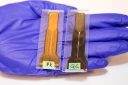
The key element of the design is NYU's high-impedance coil, which eliminates the electrodynamic interactions between the receiving components in a phased-array coil. Interestingly, the researchers achieved this goal with a relatively simple solution.
"We are using off-the-shelf coaxial cable," Cloos said. "We started by using 3D printers to make our own materials, but we quickly realized we could find materials that are commercially available and that we could use in the glove."
With no electric current created by the MR signal, the new receiver coils no longer create magnetic fields that interfere with their neighbors, thus removing the need for rigid structures to keep the coils in place. The resulting flexibility of the MRI design is "very cool," Cloos said, with the coils stitched into a cotton glove to generate images as the hand moves freely, performing different tasks.
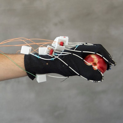
 The flexible MRI device fits like a glove and creates images from hand movements. All images courtesy of Martijn Cloos, PhD.
The glove-shaped scanner creates a 3D image that captures the entire hand and wrist. With additional coils and hardware, the researchers could extend the glove farther up the arm.
 The glove design provides a T1-weighted MR image of a hand holding a peach.
Expanded uses
So far, the NYU researchers have scanned a handful of volunteers -- no pun intended -- with this prototype. Over the next year or so, they plan to create a second version with more elements that is "more robustly engineered for more routine use," Cloos said.
"Then we will scan a much larger number of volunteers and possibly include some initial volunteer-based scans with pathology present," he said.
"There is a lot more information we can learn if we can see the hand or any other joint in different positions," Cloos said. "For example, when you are squeezing down on something, the joints may interact in a different way that we may not be able to study if they were flat and stretched out in a traditional coil."
The researchers also see expanding the use of the technology to the knee by building different-sized flexible coils that could wrap around the joint to image its movement.
"We could apply it basically across the body, and it could be a method or a key element to making more comfortable and more adaptive patient coils," Cloos added.
In addition, the researchers believe the technology could lead to more definitive guides to the anatomy of the hand, help guide surgery with images of the hand and fingers in more realistic positions, or aid in the design of better prosthetics.