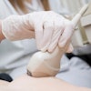
Ultrasound manufacturers are looking for ways to help their customers work faster and more efficiently, but to understand how a particular task can be done better, companies need to know exactly how it is tackled now. In the exhibition halls at ECR 2016, vendors will display equipment developed after asking sonographers and others about how they carry out routine tasks and if they can identify factors likely to cause obstructions in the hospital workflow.
On the Siemens Healthcare stand, staff are demonstrating three new additions to the Acuson range of ultrasound systems: two midrange systems, NX3 and NX3 Elite, and an upgrade to the existing premium platform, HELX Evolution with Touch Control. All offer features intended to enhance ergonomic efficiency and have been developed in consultation with people who should know how to get the most from these systems.
"Typically in most industries, a manufacturing company will look at its own technology, think how it could be improved and then develop the product. But we asked our customers what they would do if they could design their own system," said Miguel Trigueiros, director of global product marketing with Siemens' ultrasound business. "We spent three years and several million euros on this project, holding 170 workshops in five different countries where we talked to 395 sonographers. We watched them perform a range of standard tasks, listened to what they had to say, and worked together to define the challenges that they face. The feedback we received has allowed us to offer improvements which will enable them to carry out faster examinations with fewer errors, and less need for repeat exams."
The NX3 is a shared service system intended for use at general hospitals in a range of departments, notably general medicine, obstetrics/gynecology, pediatrics, and neurology. It has the largest touch-screen panel in its class, with reduced number of function buttons positioned to encourage more intuitive movements by the operator. Trigueiros estimates that a routine ob/gyn examination can be carried out with up to 28% fewer key strokes.
"As companies, we worry about the big issues, like improving image resolution and transducer quality, but these apparently minor changes are important because they make the system easier to use. We tell customers that it has been designed by them, for them," he noted.
Although Carestream is a relative newcomer to the ultrasound market, its experience in other modalities has demonstrated that its customers are a key source of new ideas. At ECR 2016, the company is unveiling its Touch Prime ultrasound system, intended for general diagnostic ultrasound imaging and developed in collaboration with customers around the world.
Touch Prime XE is the company's flagship ultrasound product, using Touch Prime Syntek to provide enhanced resolution and increase the number of images to show moving structures while optimizing image output with less noise and fewer artifacts. Improvements in imaging and Doppler provide a stable display of subtle contrast differences in tissue and can improve the ability to see small structures, according to the vendor. The Touch Prime XE unit can also achieve frame rates of over 100 Hz while maintaining image quality and functionality, as DICOM, wireless connectivity, barcode and card readers, and elastography can all be selected within the system.
Toshiba is demonstrating a range of new functions for its Aplio 500 Platinum platform, including 3D shear wave elastography, fusion imaging, and real-time 3D needle tracking.
 Leydig cell in the testis is shown using sheer-wave elastography. Image courtesy of Toshiba.
But the technology that is seen by the company as a 'game changer' is the 3D Superb Micro-Vascular Imaging (SMI) feature. This allows clinicians to see smaller vessels in and around structures like tumors, inflamed tissue, and lymph nodes, thanks to its capacity for visualizing low-velocity flow at a level far beyond the capability of conventional Doppler techniques. Significantly, SMI improves visualization of microvasculature, even without contrast agents, the company states.
Like its competitors, GE Healthcare is aware of the many problems bearing down on radiology department staff. That includes not just pressure on time and resources but also the need to maintain diagnostic standards for an ageing and increasingly obese patient population.
The firm is introducing the Logiq XDclear family, which provides a wider portfolio of probes, sophisticated anatomical models and a package of features aiming to improve clinical confidence with difficult-to-scan patients. The system offers a range of dedicated autooptimization and assistance tools to improve both productivity and the patient experience, through solutions intended to reduce the need for invasive tests and resulting patient discomfort.
 Fibrosis staging with 2D shear-wave elastography performed on the Logiq XDclear unit. Image courtesy of GE Healthcare.
This latest offering includes Logiq E9 XDclear 2.0, a new processing chain that optimizes the imaging process from the pulse of the probe to each of its pixels, delivering excellent image quality across a wide variety of cases, according to GE. The availability of extra tools such as 3D GPS Markers, MRI Auto-Registration and specialty probes will enhance its value, the company says.
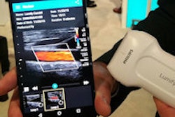
Philips Healthcare is certain to have a stream of visitors to its stand wanting to learn about Lumify, a work-in-progress. It is designed for use by emergency departments and critical care centers, as well as other clinical settings, and will operate from a compatible smart device connected to a Philips ultrasound transducer. Users will also have access to an online portal where they can manage their device and access the vendor's support, training, and IT services.
"App-based ultrasound provides valuable information to the right people at the right time. It's designed to drive a transformation in care delivery and digital health," said Vitor Rocha, chief executive of Philips' ultrasound business.
Meanwhile, Hitachi Medical Systems is launching two new members of its Arietta range of ultrasound platforms. Both the Arietta Precision and Prologue use the same core technology and electronics, but with an application-specific design for the housing. The former offers versatility for interventional and surgical procedures, the company states. Its transducer range covers minimally invasive investigations through to full open surgery, and it also has options for customized use in the operating room, with a large monitor that can be suspended from a wall or support arm and can be viewed from a distance.
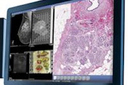
 Above: Image of the liver using the Hitachi Precision system. Below: Image of adult heart using the Hitachi Prologue ultrasound system.
The Arietta Prologue has a compact, portable design for point-of-care use. It can be cart-based or hand-carried, with an in-built battery for emergency use. It is equipped with a transducer range suitable for general abdominal examinations, musculoskeletal and vascular investigations as well as anesthesiology functions.
Fujifilm is unveiling an ultrahigh-frequency clinical ultrasound system, Vevo MD, which was developed by its North American subsidiary VisualSonics and operates at up to 70 MHz, offering the potential for unparalleled image resolution. With applications across a range of clinical areas, including neonatology, vascular, musculoskeletal imaging, and dermatology, the unit only received European regulatory approval in January 2016.
"The Vevo MD allows medical professionals to see what they have never seen before -- unparalleled image resolution down to 30 micrometers. This is less than half the size of a grain of sand," said Renaud Maloberti, general manager of Fujifilm VisualSonics.
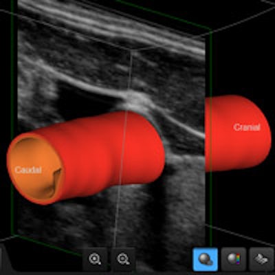
On the Samsung stand, visitors can see a demonstration of the company's latest cardiovascular ultrasound technology. Called S-3D Arterial Analysis, this function supports earlier detection of disease by executing both morphological and functional analysis of the vasculature. It enables a quantitative 3D analysis that estimates the volume of plaque within the vessel and calculates the distance to adjoining anatomical features, providing a more confident diagnosis and easier examination, the company noted.
 Screen capture demonstrates Samsung's S-3D Arterial Analysis technology for cardiovascular investigations.
Originally published in ECR Today on 2 March 2016.
Copyright © 2016 European Society of Radiology