A computer-aided detection (CAD) algorithm analyzed virtual monochromatic dual-energy CT colonography (CTC) images accurately with few false positives in a study from Massachusetts General Hospital. The researchers believe the study could pave the way to more accurate minimal-prep CTC.
The group applied its advanced CAD algorithm to CTC exams acquired with a minimal-prep protocol and virtual monochromatic dual-energy scanning. The technique detected 96% of 22 colonoscopy-confirmed lesions and 100% of all advanced lesions, with 6.1 false positives per patient for both classifications.
The system performed at least one-third better than it did on single-energy images, the group reported in a December talk at the RSNA 2014 meeting in Chicago.
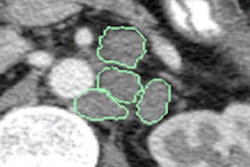
 Virtual monochromatic images reduce beam-hardening artifacts. All images courtesy of Janne Nappi, PhD.
Looking for better minimal-prep CTC
The research was conducted as part of the quest to find new tools for noncathartic CTC, which, by eliminating the uncomfortable laxative regimen, could substantially improve participation in colorectal cancer screening, said Janne Nappi, PhD, from Massachusetts General Hospital (MGH) and Harvard Medical School, in his talk.
Conventional single-source CT has limitations in noncathartic scanning, for example, in "addressing partial-volume tagging artifacts and detecting fecal tagging, because it relies on a single attenuation measurement only," he said.
 Janne Nappi, PhD, from MGH and Harvard.
The result is missed colonic lesions. More than two-thirds of all CAD misses in CTC come from unclearly tagged fecal residue (40%) or partial-volume tagging artifacts (25%), according to a previous MGH study.
But dual-energy CT, in which scans are performed at two separate energy levels, could help.
"We're investigating the use of dual-energy CT, which enables material-based detection to better identify the fecal tagging agent and soft-tissue lesions, and also to identify partial-volume fecal tagging artifacts," Nappi said. "Dual-energy CT can be used to synthesize virtual monochromatic images, and these can be used to correct for beam-hardening artifacts due to fecal tagging, and to correct for pseudoenhancement artifacts due to tagging. Ultimately, these will provide quantitatively accurate attenuation for improving the detection accuracy of computer-aided detection."
Though these so-called virtual monochromatic images can be synthesized from dual-source CT, the performance of CAD designed for use with these images is not well-understood.
The study included 66 asymptomatic screening subjects (mean age, 63 years; 47% men), all of whom underwent dual-energy CTC, according to Nappi and colleagues Hiroyuki Yoshida, PhD; Dr. Daniele Regge; Radin Nasirudin; and Peter Noel, PhD. The subjects ingested 50 mL of iodinated contrast (Gastrografin) the day before the exam (2 x 25 mL), and they had another dose (50 mL) two hours before dual-energy CTC acquisition on a dual-source scanner (Somatom Definition Flash, Siemens Healthcare).
The CT acquisitions were performed at 15 mAs for the 140-kVp scan and at 40 mAs for the 80-kVp scan. The team reconstructed dual-energy images using an iterative reconstruction dose-reduction protocol (sinogram-affirmed iterative reconstruction, or SAFIRE) with reconstruction intervals of 0.6 mm to 1.0 mm.
Multistep CAD algorithm
The CAD algorithm was designed to perform colon extraction, virtual bowel cleansing, and soft-tissue lesion detection based on material-decomposition features and the virtual monochromatic images.
Nappi and colleagues first calculated material decomposition from the images to identify typical colon materials (air, fat, soft tissue, and fecal tagging), and virtual monochromatic CTC images were reconstructed from the material-decomposition data at 125 keV to minimize image artifacts.
A support-vector machine algorithm was used to reduce false positives based on shape, texture, and material-decomposition features of the detected lesion candidates. For pilot evaluation, the per-lesion detection performance of CAD was estimated by using the leave-one-patient-out method.
Multispectral imaging features were assessed for 80-, 100-, and 140-KeV virtual monochromatic images. The data were used to assess per-lesion sensitivities and false positives for 6- to 9-mm lesions, lesions 10 mm and larger, and biopsy-confirmed neoplasms. Detection performance was compared with the performance of a similar CAD system optimized for single-source CT and with laxative-free single-source CT. Radiation dose was also assessed.
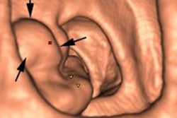
 A 16-mm flat villous adenoma was detected by the virtual monochromatic image CAD scheme, but not the single-source CAD.
The CAD algorithm's method for reducing false positives is based on conventional shape and texture features, Nappi explained. Per a 2012 paper by the group, dual-energy CT features are also employed to reveal false positives, including measurement of dual-energy index (DEI) and dual-energy ratio (DER), the partial-volume artifact index and soft-tissue index, and multispectral texture features from 80-, 100-, 120-, and 140-KeV virtual monochromatic images.
Higher sensitivity, steady false-positive rate
In all, the study revealed 18 significant biopsy-confirmed neoplasms in 18 patients, Nappi said. The patients were symptomatic, and some had undergone incomplete colonoscopy or refused conventional colonoscopy.
CAD showed 100% sensitivity at a median of three false positives per patient for neoplasms 10 mm and larger, and it detected all significant findings in the 6- to 9-mm lesion category. For neoplasms 10 mm and larger, CAD on dual-energy CT virtual monochromatic images showed a 28% increase in sensitivity (p = 0.001), compared with CAD using conventional single-energy CT, with no increase in false-positive detection. Overall, false positives were 6.1 per patient.
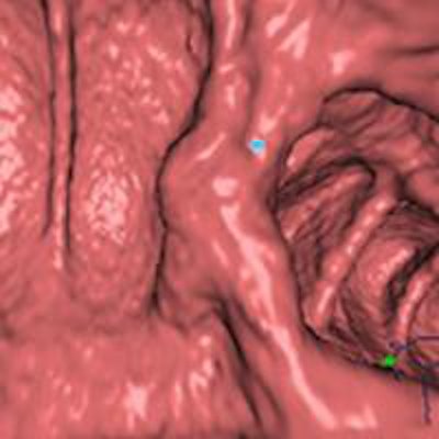
 CAD with virtual monochromatic images from dual-energy CT showed higher lesion detection than similar acquisition with conventional single-source CT.
The average CT dose index volume (CTDIvol) was 0.95 mGy, and the effective dose was 0.75 mSv per CTC scan position.
"Virtual monochromatic and material-decomposition image features can be used to yield high CAD detection performance in noncathartic dual-energy CT colonography," Nappi said. "Virtual monochromatic imaging improves the accuracy of CT attenuation measurements. Iterative reconstruction enables the use of low doses, and virtual monochromatic computer-aided detection outperforms conventional computer-aided detection."