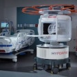
Imaging has contributed to significant advances in healthcare delivery, leading to better health outcomes and reduced costs. Technology that was once unimaginable is now the medical standard of care.
But that is just the tip of the iceberg. The next generation of imaging technologies will not only detect the location of a tumor, it will provide details about the molecular structure of the abnormal tissue within a single scan. In other words, imaging has not just obviated the need for exploratory surgery -- it may also soon render biopsies obsolete.
That's why MITA has launched the Imaging Forward campaign: to highlight the groundbreaking innovations in medical imaging technologies and the benefits of these advances on the practice of medicine. Here are some interesting facts about medical imaging you may not know.
 MITA Executive Director Gail Rodriguez, PhD.
Imaging saves lives through early detection. Low-dose lung CT finds tiny tumors the size of a grain of rice, thereby reducing lung cancer deaths by 20% compared to chest x-ray.
Imaging has virtually eliminated exploratory or unnecessary surgery. A study published in the New England Journal of Medicine found that CT reduced both the negative appendectomy rate (surgeries in which the patient turns out not to have had appendicitis) and the number of unnecessary admissions for observation, for an estimated savings of $3,540 per patient.
Medical imaging provides a safer alternative to invasive procedures. CT colonography (CTC), also known as virtual colonoscopy, uses a low-dose CT scan to spot polyps and tumors in the colon. It is faster to perform than traditional colonoscopy and avoids perforations, a possible complication of colonoscopy. A 2010 study found that 37% of those who had colon screening with CTC said that they would not have had a traditional colonoscopy at all.
Imaging provides cost-effective triage. In a study of more than 2,600 patients with mild head injury randomized to receive CT or observation, the clinical outcomes were the same: both groups of patients had good outcomes. However, the initial costs per patient were 32% lower in the CT group ($806 versus $1,183). Additionally, in a rigorous clinical trial evaluating the use of coronary CT angiography (CCTA) versus standard care for patients with chest pain and possible coronary artery disease, CCTA allowed more patients to be discharged safely from the emergency department.
During a medical emergency, imaging helps doctors make a diagnosis more quickly. Researchers at the Medical University of South Carolina found that using portable CT in patient rooms cuts the amount of time required to make images available from 50 minutes to just 20 minutes.
Imaging helps target radiation therapy. Advances in image-guided radiation therapy (IGRT) have refined cancer care, helping radiologists target tumors with great precision while limiting harm to healthy cells. This is especially useful in treating moving organs, such as the lungs, or tumors located near critical organs such as the heart. In one study, prostate cancer patients treated with IGRT fared significantly better three years after treatment than those who did not receive IGRT.
Imaging has refined cancer care. PET identifies where cancers have spread in the body and monitors the effectiveness of chemotherapy. By detecting less aggressive cancers, this powerful approach can help appropriately guide treatment. Studies have shown PET to be effective in staging certain types of lung cancer, Hodgkin's lymphoma, and colorectal cancer, and determining if cancer cells have spread to lymph nodes -- all without lifting a scalpel.
Imaging promotes high-quality healthcare. Rather than going in "blind" to place a catheter, physicians today use ultrasound to guide it precisely to the right position. Real-time ultrasound guidance for central vein catheter insertion cuts costs, decreases complications, reduces treatment time, and speeds recovery. Additionally, a study found that using CT or MRI altered treatment for 10 out of 23 stroke patients, helping to determine which patients would benefit from clot-busting drugs.
Imaging is essential to personalized healthcare. Twenty years ago, screening for breast cancer meant two things: 2D x-ray mammograms and breast physical exams. Today, not only are there more options -- including MRI to detect tumors in dense breast tissue, ultrasound, and positron emission mammography (PEM) -- it's also possible to tailor screening to the patient, rather than taking a one-size-fits-all approach. The result? Doctors find more tumors early and fewer women must suffer the follow-up screening, invasive tissue biopsy, and worry that accompanies these procedures.
We are living in what has been dubbed the "technological revolution" -- an era of unprecedented innovation in medical care. Over the past two decades, innovations in diagnostic imaging have contributed to extraordinary advances in healthcare delivery and patient outcomes. In less than a generation, imaging manufacturers have advanced the detection, diagnosis, and treatment of myriad diseases.
Imaging innovations on the horizon will allow for more targeted diagnosis and treatment, clearer images, and a wider range of cost-effective options to treat complex medical conditions. Continued investment by manufacturers is critical to ensuring physicians and patients have access to this outstanding new wave of imaging technologies.
Gail Rodriguez, PhD, is executive director of the Medical Imaging and Technology Alliance (MITA), the collective voice of medical imaging equipment, radiation therapy and radiopharmaceutical manufacturers, innovators, and product developers.


