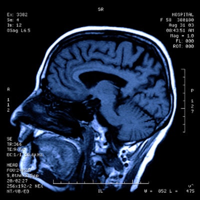
Particular brain MRI findings in some COVID-19 patients are associated with increased likelihood of the presence of the SARS-CoV-2 virus in cerebrospinal fluid, according to a study published June 9 in the Journal of Neuroimaging.
But the combination of these brain MRI results -- specifically hyperintense lesions in the brain/spine and leptomeningeal enhancement -- and SARS-CoV-2 in the spinal fluid is actually rare, suggesting that its presence is likely due to other COVID-19-induced conditions, a group led by Dr. Ariane Lewis of NYU Langone Medical Center in New York City noted.
"Although hyperintense lesions in the brain/spine and leptomeningeal enhancement are associated with the presence of SARS-CoV-2 in the cerebrospinal fluid, it is important to recognize that a positive SARS-CoV-2 [diagnostic test] in the cerebrospinal fluid is uncommon," Lewis said in a statement released by the journal. "These imaging findings are predominantly the result of inflammation, hypoxia, or ischemia, rather than infection, of the central nervous system."
Case studies have shown that central nervous system hyperintense lesions and/or leptomeningeal enhancement may appear on brain imaging in COVID-19 patients with neurological symptoms, but the cause of this has not been clear, the group noted. Clarifying the cause could improve how clinicians manage COVID-19 patients.
"These data [used in our study] may improve our understanding of the pathophysiology of these neuroimaging findings and guide decision-making about cerebrospinal fluid testing in patients with COVID-19 who have neurological symptoms," the authors wrote.
Lewis and colleagues conducted a literature review of Medline and Embase articles published between December 2019 and November 2020, gathering data to evaluate cerebrospinal fluid results in COVID-19 patients with neurological symptoms who had MRI findings that showed central nervous system hyperintense lesions not due to ischemia and/or leptomeningeal enhancement. Whether patients had SARS-CoV-2 in the cerebrospinal fluid was determined by lumbar puncture and/or polymerase chain reaction (PCR) test.
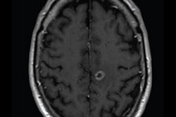
Out of 117 articles, the team identified 193 COVID-19 patients who met study criteria. Of these 193, 125 (65%) had central nervous system hyperintense lesions and were more likely to have a positive PCR test for SARS-CoV-2 in the spinal fluid (10% compared with 0% in those patients who did not have these lesions). Of 75 patients who underwent contrast MRI, 20 (27%) had leptomeningeal enhancement, and they were more likely to have a positive PCR test for SARS-CoV-2 in the spinal fluid compared with those who did not have this enhancement (25% versus 5%).
So what's the takeaway? Even though the researchers found that the association between these MRI imaging findings and SARS-CoV-2 virus in the cerebrospinal fluid is rare, the study results add to current research regarding how viruses affect the brain -- which will be helpful going forward post-COVID-19.
"Awareness of the MRI findings that can be seen in patients with evidence of SARS-CoV-2 in the cerebrospinal fluid will also be important once the pandemic is over when COVID-19 is no longer part of the differential diagnosis for every patient, as there is a characteristic MRI phenotype associated with many viral encephalitides," they concluded.


