CHICAGO - Gadolinum-enhanced MR angiography (MRA) for following up coil treatment in intracranial aneurysm patients makes the exam more costly and less efficient, according to a presentation this week at the RSNA meeting.
While MRA is widely accepted for the surveillance of these patients, "the ideal protocol for MRA has yet to be established, especially with regard to the use of intravenous gadolinium for the detection of aneurysm remnants," wrote Dr. Richard Bonsall and colleagues from the Medical University of South Carolina in Charleston in their poster presentation.
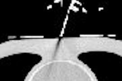
For this retrospective study, 39 patients underwent endovascular coiling and were imaged with 3D time-of-flight (TOF) MRA on a 1.5-tesla magnet. The majority of patients (51%) had aneurysms that were between 6-10 mm in size and 73.5% of the aneurysms had anterior circulation. The field-of-view (FOV) was 160 and the slice thickness was 0.5 mm. Each sequence took five minutes and 24 seconds.
Imaging was done before and after gadolinium administration. Source images, as well as maximum intensity projection (MIP) images, were examined to assess for residual flow within the coiled aneurysms and for venous contamination.
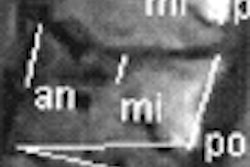
A total of 21 aneurysms were evaluated (16 anterior circulation, five posterior circulation), according to the results. Residual flow was present in 11 of the 21 aneurysms. The authors said there was no instance in which remnants were rendered visible by the gadolinium-enhanced images. In three cases, postcontrast images did enhance residual flow, but not significantly more than precontrast images.
More importantly, in five cases the contrast agent filled veins that were adjacent to the aneurysm, which mimicked residual flow, the authors noted.
"Contrast was initially proposed because it is believed that some areas of slow flow in the residual aneurysm will appear hypointense on 3-tesla TOF MRA and subsequently will be misinterpreted as aneurysm occlusion," the group wrote.
However, MRA protocols that decrease slice thickness and FOV, and have a shorter scan time, can improve imaging enough to avoid contrast altogether, they added.
Bonsall's group supports previous research that also jettisoned gadolinium-enhanced MRA in aneurysms treated with coils. French researchers from the Hospital Bretonneau in Tours also found that adding a contrast agent did not improve the ability to 3D TOF MRA to depict the presence of residual or recurrent aneurysms. They also noted that using contrast did not help visualize parent and adjacent arteries (American Journal of Neuroradiology, October 2003, Vol. 24:9, pp. 1797-1803).
By Shalmali Pal
AuntMinnie.com staff writer
November 27, 2006
Related Reading
Endovascular repair of acute abdominal aortic aneurysm less costly than surgery, September 4, 2006
Success rate low with polymer-coated platinum coils for intracranial aneurysms, April 26, 2006
Copyright © 2006 AuntMinnie.com