SINGAPORE -- New MRI techniques reveal subtle tissue alterations involved in mild traumatic brain injuries (mTBI) and provide new insights into a condition affecting millions of people worldwide, according to a leading expert.
David Wright, PhD, a neuroscientist at Monash University in Melbourne, Australia, delivered a presentation in a plenary session on the topic at the International Society for Magnetic Resonance in Medicine meeting May 6 in Singapore.
“Mild TBI is arguably the most heterogeneous of all neurological disorders. There is tremendous variability in the initial insult, from falls and sporting injuries through to explosive devices and high-speed motor vehicle accidents,” he said.
MRI is sensitive to the subtle pathophysiology that can persist beyond the resolution of symptoms and is uniquely positioned to address some of the key questions pertaining to patient management and treatment following mTBI, including for instance when it may be safe for individuals to return to pre-injury activity, Wright noted.
Yet to date, despite this promise, there is no universally accepted imaging biomarker that can characterize the full temporal progression of injury and recovery, or predict outcomes in patients, Wright said.
To that end, Wright and colleagues have conducted preclinical work to develop MRI animal models for assessing mTBI and have recently begun to see their findings validated in human clinical studies.
“Preclinical models aim to create a replicable and homogeneous injury to investigate mild TBI pathophysiology in a tightly controlled time and cost-efficient manner,” Wright said.
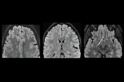
For instance, a study in rats exposed to single and then repeated mTBIs revealed via diffusion MRI – a technique used to examine the structure of axon fiber tracts in the brain – that mTBI results in long-term structural changes in the ipsilateral corpus callosum, a region of the brain consisting of white matter tracts connecting the left and right cerebral hemispheres.
“We then wanted to take this work and look at this in a clinical setting to see whether there were similar findings,” Wright said.
So, the group conducted a study of recently retired Australian professional rugby players who had histories of concussions. Compared with healthy control subjects, based on different MRI diffusion tensor imaging metrics, they saw that the ex-rugby players had reduced fractional anisotropy (FA), a biomarker of white matter microstructure, and increased diffusivity measures.

“This was nice and consistent with our preclinical study,” Wright noted.
Ultimately, these are still early days in the field, he said. Moving forward, Write noted that it is unlikely that only one imaging biomarker will characterize the full spectrum of injury and recovery in mild TBI. Instead, it will take a combination of different imaging modalities, such as perfusion imaging, chemical exchange, and saturation transfer, he noted.
“Sharing is going to be really important to boost samples and address the heterogeneity we see in mTBI as well,” Wright said.