J Thorac Imaging 1996;11(3):165-175. Atypical manifestations of pulmonary atelectasis.
Gurney JW
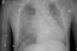
Recognizing atelectasis has always been a challenge. Atypical patterns further our knowledge of this subject. The lung has two mechanisms to help keep the lobes inflated: collateral ventilation and trapped nitrogen both tend to inflate the lungs when the airways are obstructed. Peripheral upper-lobe atelectasis resembles apical pleural fluid. Instead of collapsing superomedially, the upper lobe collapses posterolaterally, marginated by either the middle lobe or the superior segment of the lower lobe. This pattern may also be produced by segmental atelectasis of the apical-posterior segments of the upper lobe. Combined right-upper- and middle-lobe atelectasis usually stems from malignancy and violates Felson's double lesion sign. Upper-lobe atelectasis may produce a localized pneumothorax (pneumothorax ex vacuo), analogous to the vacuum joint phenomenon. Conversely, a large pneumothorax may cause torsion of an upper-lobe bronchus, leading to atelectasis. It is important to distinguish between these two conditions in order to choose the appropriate treatment-bronchoscopy in the former and chest tube drainage in the latter. Round atelectasis is a form of peripheral atelectasis that is variable in size and is thought to occur either when the lung collapses around a cleft in the presence of a pleural effusion or when shrinkage of a pleural scar pinches the adjacent lung. Round atelectasis has many features of plate atelectasis and may represent a special form of this condition.
PMID: 8784730, MUID: 96379167