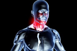
CHICAGO -- Quantitative MRI reveals how muscles in the neck are involved in primary headaches, according to research presented November 29 at the 2023 RSNA annual meeting.
In his talk, Nico Sollmann, MD, PhD, from University Hospital Ulm and University Hospital Rechts der Isar in Germany presented findings suggesting that increased T2 values of the trapezius muscles could be an objective imaging biomarker for myofascial involvement in primary headache disorders.
He said that this could help improve patient phenotyping and therapy evaluation.
Tension-type headache is the most prevalent primary headache disorder, which neck pain is tied to. However, Sollmann said no objective biomarkers exist for the myofascial involvement in primary headaches. Myofascial pain is connected to inflammation or irritation of the muscle or of the connective tissue, known as fascia, that surrounds the muscle.
The researchers wanted to study how the trapezius muscles are involved in primary headache disorders, using quantitative MRI. They also wanted to explore potential ties between muscle T2 values and the frequency of headache and neck pain.
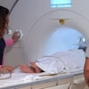
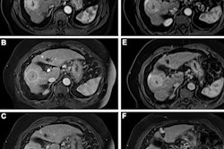
In its prospective study, the team included 50 participants. Of these, 16 had tension-type headache and 12 had tension-type headache plus migraine episodes. The groups were matched with 22 healthy controls. The study participants underwent 3D turbo spin-echo MRI and from there, the bilateral trapezius muscles were manually segmented, followed by muscle T2 extraction.
The researchers analyzed the following data points: associations between muscle T2 values and the presence of neck pain, number of days with headache, and number of myofascial trigger points as determined by manual palpation of the trapezius muscles.
They found that the group with headache plus migraine showed the highest muscle T2 values compared to the headache-only group (p < 0.001) or healthy control group (p < 0.001). For the migraine group, this included values of 31.4 ms for the right and left sides of the muscle.
 Trapezius muscle segmentations show (A) segmentation masks of the bilateral trapezius muscles (red areas) in a 25-year-old female and (B) in a 24-year-old male. Images and caption courtesy of the RSNA.
The team also reported significant ties between muscle T2 with the number of headache days (beta coefficient, 2.04; p = 0.04) and the presence of neck pain (odds ratio, 2.26; p = 0.04). It also found that the area under the curve for differentiating between healthy controls and the migraine group was 0.82, using muscle T2 as the predictor.
Finally, the researchers reported no statistically significant association between muscle T2 and the number of myofascial trigger points of the trapezius muscles.
Sollmann said that these results support the role of neck muscles in the pathophysiology of primary headaches. He added that treatments focused on the neck muscles could lead to the relief of neck pain and headache.
“Our imaging approach with delivery of an objective biomarker could facilitate therapy monitoring and patient selection for certain treatments in the near future,” he said.