VIENNA - A window into the world of radiology in France was opened wide on Friday, when three eminent researchers gave presentations on key areas of advanced imaging during the popular and well-attended European Society of Radiology (ESR) Meets France session.
In more than 20% of suspected cases of stroke, the final diagnosis is not in fact stroke, and MRI is much more sensitive than CT in diagnosing mimics of stroke. However, CT is more available, is faster, and involves fewer contraindications, according to Dr. Xavier Leclerc, a neuroradiologist at the Hôpital Roger Salengro in Lille.
 Dr. Xavier Leclerc from Lille, France.
In stroke patients, the main objectives of CT perfusion and diffusion MRI are to exclude hemorrhage, eliminate another diagnosis (i.e., mimics of stroke), detect the infarct core, evaluate the salvageable tissue (penumbra), and assess the intracranial circulation.
CT angiography (CTA) is more accurate than MR angiography (MRA) for evaluating intracranial circulation, he commented. The sensitivity and specificity of CTA is greater than 90% for detecting proximal vessel occlusion, and it is an accurate method to evaluate distal vessels and collateral circulation.
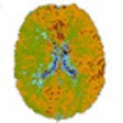
Functional imaging can be a useful tool for the radiologist, maintained Dr. Alexandre Krainik, from the department of neuroradiology and MRI at University Hospital of Grenoble. He thinks blood oxygen level dependent functional MRI (BOLD fMRI) can map functional change in perfusion and oxygenation to particular stimuli.
 Dr. Alexandre Krainik from Grenoble, France.
To make full use of the technique, however, radiologists must develop knowledge of anatomy, pathophysiology, and postprocessing, as well as mastering examination workflow. "Don't stay alone. Ask the fMRI family for help and advice," suggested Krainik.
For additional information, he urged delegates to read his electronic poster at the EPOS exhibition.
At the same session, Dr. Vincent Dousset, from the Service de Neuro-Imagerie Diagnostique et Thérapeutique at CHU Bordeaux, provided an overview of demyelinating diseases.
"In the literature, ample evidence exists to indicate that small vessel disease in the brain is the most prevalent neurological disorder ever described," he told ECR delegates. "White-matter abnormalities are almost 100% for the over 50s. Prevalence increases with age, blood pressure, diabetes, tobacco, and migraine."
 Dr. Jean-Pierre Pruvo from Lille, France.
Around 200 junior radiologists from France are attending ECR 2011. This is a very positive development, noted Dr. Jean-Pierre Pruvo, secretary general of the French Society of Radiology (SFR). SFR was founded in 1909 and has invested considerable resources in the radiology portal, www.sfrnet.org. It currently has about 8,200 members, 3,850 of whom are ESR members. A new English-language edition of the peer-reviewed Journal de Radiologie is due to be published next year.
Pruvo introduced Friday's session, along with co-moderator Dr. Yves Menu, ECR 2011 congress president.
Originally published in ECR Today March 5, 2011.
Copyright © 2011 European Society of Radiology


