Iterative reconstruction techniques for CT were once limited to dose reduction. But a new generation of iterative reconstruction algorithms is focused on image quality, processing data more rapidly and delivering more-natural looking images as well.
The additional imaging "horsepower" wielded by the new applications can be used to further reduce doses in coronary CT angiography and throughout the body -- or to ameliorate the effects of other common scanning complications.
Iterative reconstruction has benefited radiology by reducing image noise, radiation dose, and artifacts -- and the techniques can be optimized for specific clinical needs, said Jeffrey Mendel, MD, a radiologist consultant in Boston.
Iterative reconstruction "reduces the number of projections needed to reconstruct an image and provides improved dual-energy characterizations as well," Mendel said in May at the International Society for Computed Tomography (ISCT) meeting in San Francisco.
The first generation of iterative reconstruction schemes relies on a method known as accelerated simplified iterative method, Mendel said in his ISCT presentation. And although they definitely do the job in terms of dose reduction, they also produce profound change in image appearance -- "a subjective change in the look of images," as Mendel put it. "It's plastic, it's artificial, and it gets worse with higher levels of iterative weighting."
As a result, first-generation iterative techniques are typically blended with native images in concentrations no higher than 50%.
Why are these images funny-looking? It's because not all image noise is created equal, Mendel said. When noise is distributed unevenly, as early systems do, they can profoundly alter the look of CT images.
Typically, the measurement of noise (expressed in standard deviation, SD) is derived from the sum of all noise regardless of frequency, Mendel said. Images with identical overall noise can have markedly different appearances based on the frequency distribution of the noise.
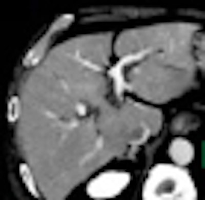
 In a CT image of the liver produced by first-generation iterative reconstruction, image shows blotchiness centrally in the liver caused by the shift in noise distribution at low frequency levels, and edge enhancement caused by the shift in image noise at high frequency levels. All images courtesy of Dr. Jeffrey Mendel.
"Think of talking in a room with a squeaky ceiling fan," Mendel said. "It's a high-pitched noise that doesn't interfere with your speech, but if somebody else in the room is talking, it makes it more challenging."
Similarly with CT images, how the noise is distributed has an important effect on how easily images can be interpreted.
The noise power spectrum, as explored by Daniele Marin, MD, and colleagues in Radiology (January 2010, Vol. 254:1, pp. 145-153), displays the frequency distribution of noise across the spatial spectrum, Mendel explained. The noise power spectrum shows the shift in the noise spectrum that is the cause of the change in appearance SD (total noise) in the area under the curve.
 Above, compared to first-generation iterative reconstruction systems, noise reduction in iDose is more evenly applied as shown in noise power spectrum graph above. Below, phantom images are similar to filtered back projection (FBP) reconstructions even at 80% dose reduction, compared to those of first-generation iterative reconstruction methods.
"These are the things that cause the plastic look that everybody is referring to, and it gets worse at higher levels of iterative weighting," Mendel said.
In contrast, the iDose algorithm (Philips Healthcare, Andover, MA) that Mendel described in his ISCT talk uses an iterative method designed to reproduce the look of full-dose filtered back projection (FBP) at a much lower dose using an iterative method. The goal is to allow use of a higher percentage of iterative reconstruction while maintaining a clinically acceptable image.
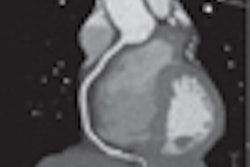
 Ultralow-dose CT acquisition in bariatric patient with iDose shows high level of detail in the heart, including detail in the mitral valve and endocardial surface (bottom) that is lost on FBP images, top.
First, the projection data itself are denoised with the application of a Poisson denoising algorithm. Then in the pixel space, iDose compares the image to a noiseless ideal anatomical model, enabling noise reduction with an appearance that is very similar to the full-dose image and does not shift the noise spectrum significantly, Mendel said.
"So even at 80% dose reduction, the images are virtually identical" to a full-dose image, even when the images are rendered, he said. In contrast, the first-generation systems show major shifts in the noise spectrum, sharpening edges and blurring other structures compared to standard images.
 iDose works in both the projection and image spaces. First, projection data are processed and projections that are data-starved are denoised using a Poisson noise distribution model. Second, in image space, noise is further reduced by fitting the data to a local structure model.
The key aspect of iDose is that it maintains the noise power spectrum (thus the appearance) of a full-dose image while utilizing high levels of dose reduction, Mendel said.
"This method allows you to use a very high level of iterative reconstruction, and what that gives us as radiologists is essentially horsepower," Mendel said. "We've talked about iterative reconstruction for dose reduction, but, remember, dose reduction may not be the only thing."
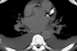
 Prospectively acquired axial coronary CT angiography (CTA) image in ultralow-dose step-and-shoot acquisition (0.25 mSv) is diagnostic with the use of advanced iterative reconstruction.
There are so many ways to reduce CT radiation dose -- reducing "padding" in prospectively gated cardiac CT scans, careful assessment of body habitus, use of automated exposure control, and more -- that further dose reductions may not be the most important factor, Mendel said.
The new method "allows us to go in other areas we made need as well: reducing artifact, bariatric imaging, and, perhaps in those patients where you really need fine detail, keeping the dose slightly higher and giving you exceptional spatial resolution," Mendel said.
 In addition to reducing dose, iterative reconstruction can address common problems such as artifacts in the shoulder region in coronal image above.
By Eric Barnes
AuntMinnie.com staff writer
August 30, 2010
Related Reading
ASIR halves VC dose without drop in image quality, June 21, 2010
MBIR aims to outshine ASIR for sharpness, CT dose reduction, May 18, 2010
Filtering software boosts CT image quality, lowers dose, May 7, 2010
ASIR cuts dose in Crohn's disease patients, December 4, 2009
Chest MDCT offers value as breast imaging tool, September 2, 2009
Copyright © 2010 AuntMinnie.com