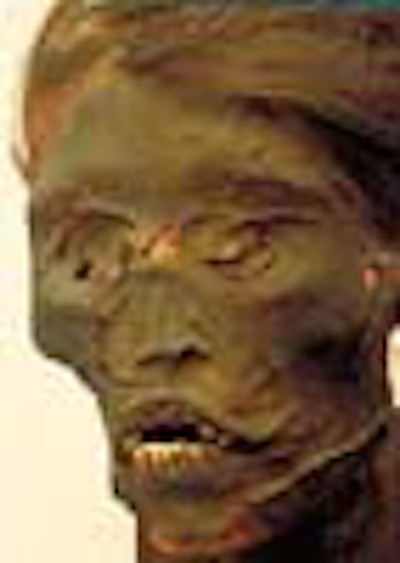
If it weren't for medical imaging, Djedmaatesankh might have been just another run-of-the-mill Egyptian mummy.
Thanks to a full-body CT scan, researchers at the Royal Ontario Museum in Canada found out a good deal about Djed without having to unwrap her from her fragile packaging: She was a middle-class, 9th century B.C. woman who lived in Thebes and most likely died after a chronically infected cyst in her upper left jaw burst open.
The connection between radiology and archaeology may seem obscure, but anthropologists increasingly are turning to imaging modalities to help them uncover the mysteries of ancient remains in a noninvasive way.
"Nondestructive techniques are really what we want," said Gerald Conlogue, RT, director of the diagnostic imaging program at Quinnipiac College in Hamden, CT. "Once you take a mummy apart, it's gone, it's done. People are more and more concerned with maintaining a certain dignity with these remains."
Conlogue and his colleagues recently founded the Bioanthropology Research Institute. Their aim is to provide on-site imaging at digs around the world using old and discarded equipment that has been re-outfitted for travel.
To that end, Conlogue is actively seeking input from the radiology community on several fronts: He's looking for donations of used equipment, volunteers to read film, and possibly even imaging professionals who are willing to travel to Peru in summer 2000 to study more than 1,000 mummies and skeletal remains.
In terms of equipment, Conlogue said he is particularly interested in the mammography units that fail to meet inspection standards under the Mammography Quality Standards Act.
"We figure that there's probably 10,000 mammography units out there that are going to become available," he said. "There's been so little contact between archaeology and anthropology and imaging folks. There's so much equipment out there now that can help change that."
When it comes to imaging mummies, aged equipment is not entirely detrimental. On one of his first field trips to Peru in the mid-1990s, Conlogue took along two Picker x-ray units from circa 1953 and 1959. The ancient machines are ideal for imaging dry, desiccated tissue, offering plenty of detail and easy use.
"Both of (the units) had the original tubes," he said. "One was a dental unit that had been in the closet in a convalescent home. It has a low penetrating power and it requires longer exposures, but since we are dealing with something that's dead, it doesn't really matter too much."
Radiation still a concern
However, one drawback of using older devices are their higher radiation levels, which may affect other readings that determine a mummy's age.
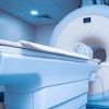
Newer technologies are also finding a place in archaeological imaging. One option is MRI, a modality recently used on a 1,000-year-old Peruvian mummy. Radiological technologist John Posh, the network MRI manager for St. Luke's Hospital Network in Muhlenberg, PA, presented his findings in a poster at the 1999 RSNA meeting.
"Until now, MRI has never been successfully used to study the ancient mysteries of mummies because researchers pre-supposed that the tissues would not contain sufficient free hydrogen to generate a signal," Posh reported.
The mummy was imaged on a GE Medical Systems 1.5-tesla EchoSpeed LX scanner. The images were generated using a T1-weighted spin-echo pulse sequence. The findings: "The sagittal T1-weighted image of the spine showed MR signal arising from the spinal canal and the intervertebral discs. The sagittal image clearly demonstrates signal arising from a structure in the posterior fossa of the skull believed to be the atrophied brain."
Posh told AuntMinnie.com that he conducted the experiment to bridge the gap between imaging and pathology.
"Medical examiners are expert at handling tissues, but they can't do the imaging," he said. "On the other hand, imaging can do the x-rays, but they aren't really equipped to handle the tissues. We have this technology and it seems a waste not to use it."
Other modalities have proved equally successful. In 1996, British explorer John Reinhard traveled to the summit of Sara Sara in Peru to retrieve the remains of two mountain dwellers. After undergoing nuclear imaging, x-rays, and CT scans, one of the bodies - dubbed Iceman - was shown to have traces of arsenic and copper in his hair. The findings are an indication that the ancient residents worked with copper far earlier than presumed.
For more information on the Bioanthropology Research Institute, contact Gerald Conlogue at [email protected].
By Shalmali Pal
AuntMinnie.com staff writer
December 20, 1999
Copyright © 1999 AuntMinnie.com