Join Dr. Steven Harms and Aurora Imaging Technology for a free, sponsor-supplied Online Symposium discussing how the AuroraCAD™ system enhances your breast MRI viewing and image processing workflow.
 Steven E. Harms, MD, FACR, Radiologist, The Breast Center of Northwest Arkansas and Medical Director, Aurora Imaging Technology. |
Join Dr. Steven Harms and Aurora Imaging Technology for a free, sponsor-supplied Online
Symposium discussing how the AuroraCAD™ system enhances your
breast MRI viewing and image processing workflow.
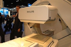
Because of the large amount of data and clinical images produced from breast MRI studies, a truly integrated and effective CAD system is a critical tool for the breast radiologist. AuroraCAD also presents simultaneous axial, sagittal and coronal views of any acquisition or post-processed image set using multi-planar reconstruction. The AuroraCAD allows for side-by-side comparison of pre-and post-contrast images, subtractions, 3D projection images, enhancement curves and more.
Aurora's user-friendly, DICOM-compatible AuroraCAD software provides the ultimate in efficiency for the physician and the technologist, which improves workflow and maximizes patient
throughput.
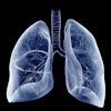
See the difference in these SpiralRODEO™ 3-D images:
 |
 |
When you're finished, feel free to post your comments and questions in the Clinical Advancements in Breast MRI discussion forum, which you can reach by clicking here.
| NOTE: | This format of an AuntMinnie Online Symposium requires Microsoft Internet Explorer 6 or later and Microsoft Media Player 9 or later. |