New techniques for coronary CT angiography (CTA) -- particularly high-pitch scanning -- are slashing radiation dose levels to provide safer scans for children without the need for sedation, according to a study presented at last week's Society of Cardiovascular Computed Tomography (SCCT) meeting in Denver.
Among several ultralow-dose techniques used by the study team from Minneapolis Heart Institute, high-pitch dual-source CTA delivered excellent image quality for a fraction of a millisievert of radiation, when heart rates could be reduced sufficiently, according to the group.
Traditionally, pediatric patients who require coronary artery imaging have undergone cardiac catheterization, an invasive procedure with a significant radiation dose that also requires sedation for all age groups, said pediatric cardiologist Dr. B. Kelly Han in an interview with AuntMinnie.com.
 |
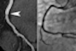
| High-pitch scan showing anomalous right coronary artery in a 17-year-old patient. Image courtesy of Dr. B. Kelly Han. |
"Children are more sensitive to radiation than adults, and they have a longer time for any radiation damage to take effect," Han said. Those with congenital heart disease typically receive many scans and a lot of radiation.
"One article estimates 20 mSv by the time they reach adolescence," mostly from catheterization procedures (approximately 3 mSv each), in addition to chest x-rays and other diagnostic studies, Han said. "Rather than look at the risk for each procedure, we're starting to look at cumulative risk over the patient's lifetime."
Newer CT scanners deliver far less radiation than previous models. However, high heart rates have been a barrier to the use of coronary CTA in children, she said. MRI is also an option, "but for small babies and for distal coronary vessels it's really not good enough yet to give us that information."
If a child has a very low likelihood of coronary artery pathology and he or she is older, the team will usually opt for MRI -- reserving CT for patients for whom MRI was not diagnostic, Han said. However, with MRI of the coronary arteries, "image quality is pretty much inversely related to age," she said. "You can see the origins pretty well but usually not the distal vessels."
Three CT protocols
For their study, Han and colleagues -- including Jana Lindberg, Dr. Robert Schwartz, Katharine Grant, PhD, and Dr. John Lesser -- reviewed all coronary CTAs performed on patients younger than 18 years at the Minneapolis Heart Institute.
Using an aggressive beta-blockade protocol to lower heart rates, combined with low-dose imaging and high-pitch scan techniques, the researchers were able to obtain excellent images of the coronary arteries in patients as young as 5 months, she said.
With the goal of comparing the image quality and the radiation dose, they examined the heart rate control with oral and IV metoprolol used for beta-blockade, and the radiation dose with the varied scan modes.
"We looked at the 76 patients who have had coronary angiograms with CT in the past four years, and categorized them by the type of scan that they got," she said. Thus, the cohort was separated by scan mode into three groups:
- Group 1: First-generation, dual-source CT scanner (Somatom Definition, Siemens Healthcare) with retrospective electrocardiogram (ECG) gating (spiral scan mode)
- Group 2: Second-generation, dual-source CT scanner (Somatom Definition Flash, Siemens) with prospective ECG gating (sequential scan mode)
- Group 3: Second-generation, dual-source CT using prospective ECG gating with high pitch (flash-scan mode)
The researchers compared age, heart rate, body surface area, radiation dose estimates, and image quality between the three groups. They performed 76 scans in patients from 3 days to 18 years of age. Image quality was subjectively rated on a scale of 1 to 4, with 4 being optimal.
"The importance of getting the heart rate down is that the percentage of the RR interval that you need to cover varies depending on your heart rate," she said. Therefore, for heart rates of 65 to 70 beats per minute, about 40% of the RR interval needs to be covered, whereas a single diastolic interval is sufficient for a slower heart rate.
The differences in radiation dose between the three scan groups were statistically significant, and high image quality was maintained between groups despite the decreased radiation exposure (mean 0.85 mSv overall).
"That's about 25% of the dose of cath for all studies of CT angiography," she said.
- The mean radiation dose for group 1 (spiral mode, retrospective gating) was 1.8 mSv, and image quality was 3.5 (22 scans; mean age, 10 years).
- The mean radiation dose for group 2 (prospective gating, sequential mode) was 1.01 mSv, and image quality was 3.3 (35 scans; mean age, 9 years).
- The mean radiation dose for group 3 (prospective gating, high pitch) was 0.035 mSv, and image quality was 3.4 (19 scans; mean age, 11 years).
There was no statistically significant difference in age, body surface area, scan heart rate, or image quality among the groups. A total of 17 patients underwent subsequent surgical intervention, and surgical findings correlated with coronary CTA in every case, she said.
 |
| Pediatric coronary CTA radiation dose estimates. Chart courtesy of Dr. B. Kelly Han. |
"With the high-pitch scans, there was an increase in noise, but it didn't affect diagnostic confidence," Han said. "We were able to see the entire coronary artery tree in patients as young as 5 months of age," and all scans were diagnostic, she said.
Coronary CTA performed on newer CT scanners using aggressive dose-reduction techniques delivers far less radiation than a catheter-based angiogram, she said. It's also noninvasive and requires no sedation for patients 7 years and older.
"I think you can do a test for less risk that maintains the diagnostic quality," Han said. "The other interesting thing is that in patients with Kawasaki's disease who have had aneurysms and stenoses, we're also finding plaque and calcium that you wouldn't find with any other modality."
For young patients with congenital abnormalities, low-dose CT provides excellent definition of structure with much less radiation, with the ability to see plaque and calcifications in certain patient subsets that would not otherwise be visible, she said.