A computer-aided detection (CAD) system can help detect early resectable lung cancer, according to an article published online in the Journal of Digital Imaging.
To evaluate a commercially available CAD system on operable T1 lung cancer cases using digital chest radiography equipment, a research team from Kyushu University in Fukuoka, Japan; Mitsubishi Space Software in Amagasaki, Japan; and the University of Chicago in Illinois studied 50 consecutive patients undergoing surgery for primary lung cancer and 50 normal cases.
Chest radiography exams were obtained using either a FCR 5501D computed radiography system (Fuji Photo Film, Tokyo), a CDXI-11 digital radiography system (Canon, Tokyo), or a Thorax FD DR system (Siemens Medical Solutions, Erlangen, Germany). Eight radiologists (four chest radiologists and four residents) interpreted soft-copy systems using a display system both with and without CAD (EpiSight/XR, Mitsubishi Space Software) output (JDI, June 14, 2006).
The cases were presented in random order and in the same sequence for all observers. Each observer interpreted all images in one session, according to the authors.
The radiographs were first shown for conventional interpretation, with the observers marking their confidence level by marking on a bar on the lower part of the monitor screen. When observers diagnosed cases as positives, they recorded the locations of lesions on paper hard copies as a numeral 1 with a red pencil.
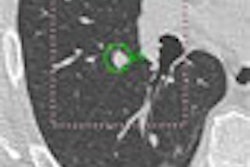
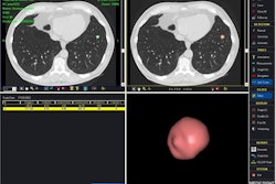
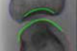
The CAD output image was then displayed on another monitor. The observers viewed the CAD output image and the radiographs, and marked their confidence level on the same bar. When observers diagnosed cases as positives and changed the location of a nodule, the new location of the lesion was recorded on hard copies as 2 without erasing numeral 1, according to the research team.
CAD detected lung cancer in 37 of the 50 (74%) patients, with a false-positive rate of 2.28 (114/50) false positives per case for normal cases. The mean area under the ROC curve increased significantly from 0.896 without CAD output to 0.923 with CAD output (p = 0.018). CAD changed false negatives without CAD to true positives in 19 of 31 (61%) of cases.
The team also found improvement in the localization of tumors in 1.5 cases, on average, for radiology residents. The group measured considerable changes from false-positive to true-negative findings for residents (10/22, 45%). Also, the ratio of beneficially changed cases to detrimentally changed cases was higher for chest radiologists (9:2) than for residents (22:13), according to the researchers.
The study team attributed performance improvement from CAD mainly due to changes of 61% false negatives without CAD to true positives.
"Improvement in determining the location of tumor was observed in 1.5 cases on average for the residents, but there was no case for which the location of the tumor was changed by the chest radiologists," the authors concluded. "This CAD system for digital chest radiographs is useful in assisting radiologists for detection of early resectable lung cancer."
By Erik L. Ridley
AuntMinnie.com staff writer
June 22, 2006
Related Reading
NCI CAD database program gathers momentum, March 16, 2006
Chest x-ray CAD reaches reimbursement milestone, November 22, 2005
X-ray CAD finds bone defects in rheumatoid arthritis, May 18, 2004
Copyright © 2006 AuntMinnie.com